Download
1 / 26
260 likes | 555 Views
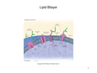
Multiscale Modeling of Lipid Bilayer Interactions with Solid Substrates. David R. Heine, Aravind R. Rammohan, and Jitendra Balakrishnan October 23 rd , 2008 RPI High Performance Computing Conference. Outline. Background structure of lipid bilayers applications of supported lipid bilayers

E N D
Multiscale Modeling of Lipid Bilayer Interactions with Solid Substrates David R. Heine, Aravind R. Rammohan, and Jitendra Balakrishnan October 23rd, 2008 RPI High Performance Computing Conference
Outline • Background • structure of lipid bilayers • applications of supported lipid bilayers • Modeling challenges • Atomistic modeling • Mesoscale modeling • Experimental work • Conclusions
Technological Relevance of Supported Lipid Bilayers • SLBs are important for various biotech applications • Biological research • Model systems to study the properties of cell membranes • Stable, immobilized base for research on membrane moieties • Biosensors for the activity of various biological species • Cell attachment surfaces • Pharmaceutical research • Investigation of membrane receptor drug targets • Membrane microarrays: High throughput screening for drug discovery • How does bilayer-substrate interaction affect bilayer behavior?
Supported Lipid Bilayers at Corning • Applications: Membrane-protein microarrays for pharmaceutical drug discovery • Substrate texture is important in the adhesion and conformation of bilayers on the surface • Crucial for the biological functionality of bilayers • Objective: Quantify the effect of substrate topography and chemical composition on bilayer conformation and dynamics
Time Scales Bond Vibrations: fs Lateral Diffusion Time: 4 ps Peristaltic Modes: 1-10 ns Undulatory Modes 0.1 ns – 0.1 ms Membrane Fusion: 1-10 s Bilayer Length & Time Scales • Bilayer dynamics vary over large length and time scales, suggesting a multiscale approach. Length Scales Stokes Radius: 2.4 nm Bilayer Thickness: 4 nm Area per lipid: 60 +/- 2 Å2 Undulations: 4 Å – 0.25 mm
Multiscale Approach • Atomistic model • capture local structure and short term dynamics • Mesoscale model • capture longer length and time scales • sufficient to look at interaction with rough surfaces
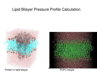
lipid water substrate Atomistic Model • The bilayer is composed of 72 DPPC lipid molecules described in full atomistic detail using the CHARMM potential • Water uses the flexible SPC model to allow for bond angle variations near the substrate • The substrate is the [100] face of a-quartz with lateral dimensions of 49 x 49 Å described by the ClayFF potential
Water Lipids Upper leaflet Bilayer Lower leaflet Water Substrate Simulation Technique • System is periodic in x and y directions with a repulsive wall above the water surface in the z direction • NVT ensemble must be used since pressure control is prohibited by the solid substrate • Temperature is maintained at 323K with a Nose-Hoover thermostat • Total energy and force on the bilayer are extracted during the simulation. Heine et al. Molecular Simulations, 2007, 33(4-5), pp.391-397.
Simulation Technique • System is periodic in x and y directions with a repulsive wall above the water surface in the z direction • NVT ensemble must be used since pressure control is prohibited by the solid substrate • Temperature is maintained at 323K with a Nose-Hoover thermostat • Total energy and force on the bilayer are extracted during the simulation. Heine et al. Molecular Simulations, 2007, 33(4-5), pp.391-397.
Comparison with Experimental Measurements Bilayer-Substrate Interaction Energy from Simulations Simulations show an energy minimum at a separation of 3 to 3.5 nm SFA Measurements Between Substrate and Bilayer Experimental measurements show a repulsion starting around 4 nm and pullout at 3 nm separations courtesy J. Israelachvili, UCSB
Lower monolayer is compressed in the vicinity of substrate Upper monolayer seems relatively unaffected Bilayer structure near the substrate
Reduction in lateral diffusivity observed, compared to free bilayers Bulk simulations match diffusivity of free bilayers Suppression of transverse fluctuations near substrate inhibit a key mechanism for lateral diffusion Effect of substrate on lateral lipid diffusion Transverse lipid motion enables lateral diffusion Substrate reduces transverse motion & reduces diffusivity Experimental value For free bilayers
Atomistic Simulation Results • MD simulations show bilayer-substrate equilibrium separation of 3 – 3.5 nm, in agreement with SFA experiments • Lateral diffusion of the lipid head groups decreases as the bilayer approaches the substrate • Suppression of transverse fluctuations may be responsible for reduced lateral diffusion
Membrane Continuum solvent Substrate Mesoscopic Model • Conservative force • Elastic stretching of bilayer • Bending modes of bilayer • Surface interactions • Other (electrostatic, etc.) • Dissipative force • Formulation based on Newtonian solvent viscosity • Random force • Formulation based on fluctuation-dissipation theorem
Continuum representation to study large length and time scales 1 mm2, 1 ms Allows study of bilayer behavior on textured substrates Dynamic model that includes effect of solvent and environment Mesoscopic Modeling of Supported Lipid Bilayers All dimensions in nanometers z axis not to scale
Mesoscopic Model Results Substrate topography contours Membrane topography contours
Membrane Coating Membrane spanning Maximum Separation Minimum Separation Mesoscopic Model Results
Mesoscopic Model Results • Allows study of bilayer on micron and microsecond scales • Minimum surface roughness of 4-5 nm required for membrane spanning conformation • Spanning configuration important for maintaining bilayer mobility
AFM measurementsSpreading of Bilayer on Synthetic Substrates AFM image & measurements courtesy Sergiy Minko, Clarkson University Ref: Nanoletters, 2008, 8(3), 941-944
AFM measurementsSmoothening of membrane on rough substrates AFM image & measurements courtesy Sergiy Minko, Clarkson University
BILAYER ~ 5 nm Maximum Separation Minimum Separation SUBSTRATE Lipid membrane conformationNumerical and Experimental Results • Model shows membrane coating up to about 4-5 nm • AFM images show membrane coating 5 nm particles Macroscopic model predictions AFM images courtesy Sergiy Minko, Clarkson U. Roiter et al. Nanoletters 8, 941 (2008)
Conclusions • MD simulations show bilayer-substrate separation of 3 – 3.5 nm, in agreement with SFA experiments • MD simulations show reduced lateral diffusion in lipids as the bilayer approaches the substrate • Mesoscopic model shows membranes coat particles up to 4 – 5 nm in diameter, in agreement with AFM observations • Larger surface features are needed to achieve separation between bilayer and substrate • High-performance computing has opened up new approaches for understanding biomolecule-substrate interactions, which aids design • There is still plenty of room to grow as these models are still restricted in terms of size, timescale, and complexity
Acknowledgements • Professor Sergiy Minko & his group at Clarkson U. • Professor Jacob Israelachvili & his group at U. C. Santa Barbara
Lipid Behavior on Nanoparticles • Bilayer conforms to Nanoparticles < 1.2 nm • Bilayer undergoes structural re-arrangement involving formation of holes between 1.2 – 22 nm • Beyond 22 nm bilayer envelops the particle Ref: Nanoletters, 2008, 8(3), 941-944