Download
1 / 58
610 likes | 1.08k Views
Thoracic Trauma. Adapted from Dave Lloyd, MD PCP Care January 28, 2012. Introduction to Thoracic Injury. Vital Structures Heart, Great Vessels, Esophagus, Tracheobronchial Tree, & Lungs 25% of MVC deaths are due to thoracic trauma 12,000 annually in US

E N D
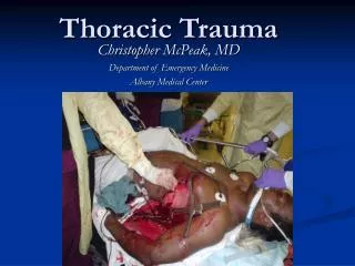
Thoracic Trauma Adapted from Dave Lloyd, MD PCP Care January 28, 2012
Introduction to Thoracic Injury • Vital Structures • Heart, Great Vessels, Esophagus, Tracheobronchial Tree, & Lungs • 25% of MVC deaths are due to thoracic trauma • 12,000 annually in US • Abdominal injuries are common with chest trauma. • Prevention Focus • Gun Control Legislation • Improved motor vehicle restraint systems • Passive Restraint Systems • Airbags
Anatomy and Physiology of the Thorax • Thoracic Skeleton • 12 Pair of C-shaped ribs • Ribs 1-7: Join at sternum with cartilage end-points • Ribs 8-10: Join sternum with combined cartilage at 7th rib • Ribs 11-12: No anterior attachment • Sternum • Manubrium • Joins to clavicle and 1st rib • Jugular Notch • Body • Sternal angle (Angle of Louis) • Junction of the manubrium with the sternal body • Attachment of 2nd rib • Xiphoid process • Distal portion of sternum
Anatomy and Physiology of the Thorax • Thoracic Skeleton • Topographical Thoracic Reference Lines • Midclavicular line • Anterior axillary line • Mid-axillary line • Posterior axillary line • Intercostal space • Artery, Vein and Nerve on inferior margin of each rib • Thoracic Inlet • Superior opening of the thorax • Curvature of 1st rib with associated structures • Thoracic Outlet • Inferior opening of the thorax • 12th rib and associated structures & Xiphisternal joint
Anatomy and Physiology of the Thorax • Diaphragm • Muscular, dome-like structure • Separates abdomen from the thoracic cavity • Affixed to the lower border of the rib cage • Central and superior margin extends to the level of the 4th rib anteriorly and 6th rib posteriorly • Major muscle of respiration • Draws downward during inspiration • Moves upward during exhalation
Anatomy and Physiology of the Thorax • Associated Musculature • Shoulder girdle • Muscles of respiration • Diaphragm • Intercostal muscles • Contract to elevate the ribs and increase thoracic diameter • Increase depth of respiration • Sternocleidomastoid • Raise upper rib and sternum
Anatomy and Physiology of the Thorax • Physiology of Respiration • Changing pressure assists: • Venous return to heart • Pumping blood to systemic circulation • Inhalation • Diaphragm contracts and flattens • Intercostals contract expanding ribcage • Thorax volume increases • Less internal pressure than atmospheric • Air enters lungs • Exhalation • Musculature relaxes • Diaphragm & intercostals return to normal • Greater internal pressure than atmospheric • Air exits lungs
Anatomy and Physiology of the Thorax • Trachea, Bronchi & Lungs • Trachea • Hollow & cartilage supported structure • Bronchi • Right & left extend for 3 centimeters • Enters lungs at Pulmonary Hilum • Also where pulmonary arteries & veins enter • Further subdivide and terminate as alveoli • Basic unit of structure & function in the lungs • Single cell membrane • External versus Internal Respiration • Lungs • Right = 3 lobes • Left = 2 lobes
Anatomy and Physiology of the Thorax • Trachea, Bronchi & Lungs • Pleura • Visceral Pleura • Cover lungs • Parietal Pleura • Lines inside of thoracic cavity • Pleural Space • POTENTIAL SPACE • Air in Space = PNEUMOTHORAX • Blood in Space = HEMOTHORAX • Serous (pleural) fluid within • Lubricates & permits ease of expansion
Anatomy and Physiology of the Thorax • Mediastinum • Central space within thoracic cavity • Boundaries • Lateral: Lungs • Inferior: Diaphragm • Superior: Thoracic outlet • Structures • Heart • Great Vessels • Esophagus • Trachea • Nerves • Vagus • Phrenic • Thoracic Duct
Anatomy and Physiology of the Thorax • Heart • Chambers • Valves • Vessels • External Vessels • Coronary Arteries • Contraction Cycle • Systole • Diastole • Filling of the coronary arteries occur
Anatomy and Physiology of the Thorax • Heart • General Structure • Pericardium • Surrounds heart • Visceral • Parietal • Serous • 35-50 ml fluid • Epicardium • Outer Layer • Myocardium • Muscular layer • Endocardium • Innermost layer • Nervous Structure • SA Node • Right Atrium • Intra-atrial Pathways • AV Node • AV Junction • Bundle of His • Left & Right Bundle Branches • Purkinje Fibers
Anatomy and Physiology of the Thorax • Great Vessels • Aorta • Fixed at three sites • Annulus • Attaches to heart • Ligamentum Arteriosum • Near bifurcation of pulmonary artery • Aortic hiatus • Passes through diaphragm • Superior Vena Cava • Inferior Vena Cava • Pulmonary Arteries • Pulmonary Veins • Esophagus • Enters at thoracic inlet • Posterior to trachea • Exits at esophageal hiatus
Pathophysiology of Thoracic Trauma • Blunt Trauma • Results from kinetic energy forces • Subdivision Mechanisms • Blast • Pressure wave causes tissue disruption • Tear blood vessels & disrupt alveolar tissue • Disruption of tracheobronchial tree • Traumatic diaphragm rupture • Crush (Compression) • Body is compressed between an object and a hard surface • Direct injury of chest wall and internal structures • Deceleration • Body in motion strikes a fixed object • Blunt trauma to chest wall • Internal structures continue in motion • Ligamentum Arteriosum shears aorta • Age Factors • Pediatric Thorax: More cartilage = Absorbs forces • Geriatric Thorax: Calcification & osteoporosis = More fractures
Pathophysiology of Thoracic Trauma • Penetrating Trauma • Low Energy • Arrows, knives, handguns • Injury caused by direct contact and cavitation • High Energy • Military, hunting rifles & high powered hand guns • Extensive injury due to high pressure cavitation Trauma.org
Pathophysiology of Thoracic Trauma • Penetrating Injuries (cont.) • Shotgun • Injury severity based upon the distance between the victim and shotgun & caliber of shot • Type I: >7 meters from the weapon • Soft tissue injury • Type II: 3-7 meters from weapon • Penetration into deep fascia and some internal organs • Type III: <3 meters from weapon • Massive tissue destruction
Injuries Associated with Penetrating Thoracic Trauma • Closed pneumothorax • Open pneumothorax (including sucking chest wound) • Tension pneumothorax • Pneumomediastinum • Hemothorax • Hemopneumothorax • Laceration of vascular structures • Tracheobronchial tree lacerations • Esophageal lacerations • Penetrating cardiac injuries • Pericardial tamponade • Spinal cord injuries • Diaphragm trauma • Intra-abdominal penetration with associated organ injury
Pathophysiology of Thoracic Trauma Chest Wall Injuries • Contusion • Most Common result of blunt injury • Signs & Symptoms • Erythema • Ecchymosis • DYSPNEA • PAIN on breathing • Limited breath sounds • HYPOVENTILATION • BIGGEST CONCERN = “HURTS TO BREATHE” • Crepitus • Paradoxical chest wall motion
Pathophysiology of Thoracic Trauma Chest Wall Injuries • Rib Fractures • >50% of significant chest trauma cases due to blunt trauma • Compressional forces flex and fracture ribs at weakest points • Ribs 1-3 requires great force to fracture • Possible underlying lung injury • Ribs 4-9 are most commonly fractured • Ribs 9-12 less likely to be fractured • Transmit energy of trauma to internal organs • If fractured, suspect liver and spleen injury • Hypoventilation is COMMON due to PAIN
Pathophysiology of Thoracic Trauma Chest Wall Injuries • Sternal Fracture & Dislocation • Associated with severe blunt anterior trauma • Typical MOI • Direct Blow (i.e. Steering wheel) • Incidence: 5-8% • Mortality: 25-45% • Myocardial contusion • Pericardial tamponade • Cardiac rupture • Pulmonary contusion • Dislocation uncommon but same MOI as fracture • Tracheal depression if posterior
Pathophysiology of Thoracic Trauma Chest Wall Injuries • Flail Chest • Segment of the chest that becomes free to move with the pressure changes of respiration • Three or more adjacent rib fracture in two or more places • Serious chest wall injury with underlying pulmonary injury • Reduces volume of respiration • Adds to increased mortality • Paradoxical flail segment movement • Positive pressure ventilation can restore tidal volume
Pathophysiology of Thoracic Trauma Pulmonary Injuries • Simple Pneumothorax • AKA: Closed Pneumothorax • Progresses into Tension Pneumothorax • Occurs when lung tissue is disrupted and air leaks into the pleural space • Progressive Pathology • Air accumulates in pleural space • Lung collapses • Alveoli collapse (atelectasis) • Reduced oxygen and carbon dioxide exchange • Ventilation/Perfusion Mismatch • Increased ventilation but no alveolar perfusion • Reduced respiratory efficiency results in HYPOXIA • Typical MOI: “Paper Bag Syndrome”
Pathophysiology of Thoracic Trauma Pulmonary Injuries • Open Pneumothorax • Free passage of air between atmosphere and pleural space • Air replaces lung tissue • Mediastinum shifts to uninjured side • Air will be drawn through wound if wound is 2/3 diameter of the trachea or larger • Signs & Symptoms • Penetrating chest trauma • Sucking chest wound • Frothy blood at wound site • Severe Dyspnea • Hypovolemia
Pathophysiology of Thoracic Trauma Pulmonary Injuries • Tension Pneumothorax • Buildup of air under pressure in the thorax. • Excessive pressure reduces effectiveness of respiration • Air is unable to escape from inside the pleural space • Progression of Simple or Open Pneumothorax
Pathophysiology of Thoracic Trauma Pulmonary InjuriesTension Pneumothorax Signs & Symptoms • Dyspnea • Tachypnea at first • Progressive ventilation/perfusion mismatch • Atelectasis on uninjured side • Hypoxemia • Hyperinflation of injured side of chest • Hyperresonance of injured side of chest • Diminished then absent breath sounds on injured side • Cyanosis • Diaphoresis • AMS • JVD • Hypotension • Hypovolemia • Tracheal Shifting • LATE SIGN
Pathophysiology of Thoracic Trauma Pulmonary Injuries • Hemothorax • Accumulation of blood in the pleural space • Serious hemorrhage may accumulate 1,500 mL of blood • Mortality rate of 75% • Each side of thorax may hold up to 3,000 mL • Blood loss in thorax causes a decrease in tidal volume • Ventilation/Perfusion Mismatch & Shock • Typically accompanies pneumothorax • Hemopneumothorax
Pathophysiology of Thoracic Trauma Pulmonary InjuriesHemothorax Signs & Symptoms • Blunt or penetrating chest trauma • Shock • Dyspnea • Tachycardia • Tachypnea • Diaphoresis • Hypotension • Dull to percussion over injured side
Pathophysiology of Thoracic Trauma Pulmonary Injuries • Pulmonary Contusion • Soft tissue contusion of the lung • 30-75% of patients with significant blunt chest trauma • Frequently associated with rib fracture • Typical MOI • Deceleration • Chest impact on steering wheel • Bullet Cavitation • High velocity ammunition • Microhemorrhage may account for 1- 1 ½ L of blood loss in alveolar tissue • Progressive deterioration of ventilatory status • Hemoptysis typically present
Pathophysiology of Thoracic Trauma Cardiovascular Injuries • Myocardial Contusion • Occurs in 76% of patients with severe blunt chest trauma • Right Atrium and Ventricle is commonly injured • Injury may reduce strength of cardiac contractions • Reduced cardiac output • Electrical Disturbances due to irritability of damaged myocardial cells • Progressive Problems • Hematoma • Hemoperitoneum • Myocardial necrosis • Dysrhythmias • CHF & or Cardiogenic shock
Pathophysiology of Thoracic Trauma Cardiovascular InjuriesMyocardial Contusion Signs & Symptoms • Bruising of chest wall • Tachycardia and/or irregular rhythm • Retrosternal pain similar to MI • Associated injuries • Rib/Sternal fractures • Chest pain unrelieved by oxygen • May be relieved with rest • THIS IS TRAUMA-RELATED PAIN • Similar signs and symptoms of medical chest pain
Pathophysiology of Thoracic Trauma Cardiovascular Injuries • Pericardial Tamponade • Restriction to cardiac filling caused by blood or other fluid within the pericardium • Occurs in <2% of all serious chest trauma • However, very high mortality • Results from tear in the coronary artery or penetration of myocardium • Blood seeps into pericardium and is unable to escape • 200-300 ml of blood can restrict effectiveness of cardiac contractions • Removing as little as 20 ml can provide relief
Pathophysiology of Thoracic Trauma Cardiovascular InjuriesPericardial Tamponade Signs & Symptoms • Dyspnea • Possible cyanosis • Beck’s Triad • JVD • Distant heart tones • Hypotension or narrowing pulse pressure • Weak, thready pulse • Shock • Kussmaul’s sign • Decrease or absence of JVD during inspiration • Pulsus Paradoxus • Drop in SBP >10 during inspiration • Due to increase in CO2 during inspiration • Electrical Alterans • P, QRS, & T amplitude changes in every other cardiac cycle • PEA
Pathophysiology of Thoracic Trauma Cardiovascular Injuries • Myocardial Aneurysm or Rupture • Occurs almost exclusively with extreme blunt thoracic trauma • Secondary due to necrosis resulting from MI • Signs & Symptoms • Severe rib or sternal fracture • Possible signs and symptoms of cardiac tamponade • If affects valves only • Signs & symptoms of right or left heart failure • Absence of vital signs
Pathophysiology of Thoracic Trauma Cardiovascular Injuries • Traumatic Aneurysm or Aortic Rupture • Aorta most commonly injured in severe blunt or penetrating trauma • 85-95% mortality • Typically patients will survive the initial injury insult • 30% mortality in 6 hrs • 50% mortality in 24 hrs • 70% mortality in 1 week • Injury may be confined to areas of aorta attachment • Signs & Symptoms • Rapid and deterioration of vitals • Pulse deficit between right and left upper or lower extremities
Pathophysiology of Thoracic Trauma Cardiovascular Injuries • Other Vascular Injuries • Rupture or laceration • Superior Vena Cava • Inferior Vena Cava • General Thoracic Vasculature • Blood Localizing in Mediastinum • Compression of: • Great vessels • Myocardium • Esophagus • General Signs & Symptoms • Penetrating Trauma • Hypovolemia & Shock • Hemothorax or hemomediastinum
Pathophysiology of Thoracic Trauma Other Thoracic Injuries • Traumatic Esophageal Rupture • Rare complication of blunt thoracic trauma • 30% mortality • Contents in esophagus/stomach may move into mediastinum • Serious Infection occurs • Chemical irritation • Damage to mediastinal structures • Air enters mediastinum • Subcutaneous emphysema and penetrating trauma present
Pathophysiology of Thoracic Trauma Other Thoracic Injuries • Tracheobronchial Injury • MOI • Blunt trauma • Penetrating trauma • 50% of patients with injury die within 1 hr of injury • Disruption can occur anywhere in tracheobronchial tree • Signs & Symptoms • Dyspnea • Cyanosis • Hemoptysis • Massive subcutaneous emphysema • Suspect/Evaluate for other closed chest trauma
Pathophysiology of Thoracic Trauma Other Thoracic Injuries • Traumatic Asphyxia • Results from severe compressive forces applied to the thorax • Causes backwards flow of blood from right side of heart into superior vena cava and the upper extremities • Signs & Symptoms • Head & Neck become engorged with blood • Skin becomes deep red, purple, or blue • NOT RESPIRATORY RELATED • JVD • Hypotension, Hypoxemia, Shock • Face and tongue swollen • Bulging eyes with conjunctival hemorrhage
Assessment of the Thoracic Trauma Patient • Scene Size-up • Initial Assessment • Rapid Trauma Assessment • Observe • JVD, SQ Emphysema, Expansion of chest • Question • Palpate • Auscultate • Percuss • Blunt Trauma Assessment • Penetrating Trauma Assessment • Ongoing Assessment
Management of the Chest Injury PatientGeneral Management • Ensure ABC’s • High flow O2 via NRB • Intubate if indicated • Consider RSI • Consider overdrive ventilation • If tidal volume less than 6,000 mL • BVM at a rate of 12-16 • May be beneficial for chest contusion and rib fractures • Promotes oxygen perfusion of alveoli and prevents atelectasis • Anticipate Myocardial Compromise • Shock Management • Consider PASG • Only in blunt chest trauma with SP <60 mm Hg • Fluid Bolus: 20 mL/kg • AUSCULTATE! AUSCULATE! AUSCULATE!