Download
1 / 1
20 likes | 378 Views
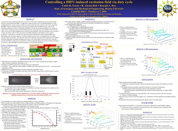
Acrylamide phantom with bovine serum albumin (HIFU source on right). “Cigar- shaped” lesion. “Tadpole- shaped” lesion. Prefocal PCD. Focal PCD. R2. Fundamental Science. R1. R3. Validating TestBEDs. Bio-Med. Enviro-Civil. HIFU on. S2. S3. S4. S1. S5.

E N D
Acrylamide phantom with bovine serum albumin (HIFU source on right) “Cigar- shaped” lesion “Tadpole- shaped” lesion Prefocal PCD Focal PCD R2 FundamentalScience R1 R3 ValidatingTestBEDs Bio-Med Enviro-Civil HIFU on S2 S3 S4 S1 S5 Controlling a HIFU-induced cavitation field via duty cycleCaleb H. Farny • R. Glynn Holt • Ronald A. RoyDept. of Aerospace and Mechanical Engineering, Boston UniversityCenSSIS RICC, October 6-7, 2005Work supported by the US Army and the Center for Subsurface Sensing and ImagingSystems via NSF ERC award number EEC-9986821. ABSTRACT Cavitation has been implicated in the lack of control over the shape of thermal lesions generated by high-intensity focused ultrasound (HIFU). Employing a single focused, passive broadband transducer in agar-graphite phantoms, we have shown a decline in acoustic emissions from cavitation at the focus, suggesting that HIFU energy is shielded from the focal region, possibly by prefocal bubble activity. Our recent modeling results show, however, that bubble shielding is not the only mechanism behind such a change in signal. As the temperature increases the broadband acoustic emissions from an air bubble in water decrease, and so a decrease in signal amplitude from cavitation events should be expected as heating occurs. In order to evaluate the relative effects of the temperature and bubble shielding on the bubble activity, we have positioned a second passive transducer at various positions in the prefocal region along the HIFU axis. Depending on the insonation pressure a decline in signal from the focus is accompanied by an eventual increase in prefocal pressure. The timescale of the focal signal decrease and prefocal signal increase suggests that both temperature and bubble shielding effects play a role in the bubble activity at the focus, and may provide information on how best to monitor the cavitation signal and ultimately provide feedback information necessary to control the HIFU insonation parameters to avoid bubble shielding. • EXPERIMENT • The rapid "inertial" collapse of bubble produces broadband emissions (“cavitation activity”).5 • Cavitation can be detected by passively listening to broadband noise emissions. • Key instrumentation elements: • 1.1 MHz focused HIFU transducer; • Two 15 MHz passive cavitation detectors (PCD): • One PCD confocal with the HIFU transducer • One PCD positioned along the prefocal region, perpendicular to HIFU axis • Agar-graphite tissue-mimicking phantom • Prefocal PCD moved in 1 mm increments between the focus and 5 mm in front of focus (in between experiments). • Three peak negative pressures: 2, 2.6, 3 MPa. • Compare cavitation activity at focus with activity at prefocal positions as a function of time and pressure. RESULTS: 2.6 MPa focal pressure • There is a rapid decrease in cavitation emissions at the focus, 1, 2 mm. • Amplitude at the focus is higher. • Cavitation emissions increase over time at 3 and 4 mm in front of the focus. • No activity 5 mm prefocal. EXPERIMENTAL SETUP Focal region CONTACT INFORMATION: Prof. Ronald A. Roy Prof. R. Glynn Holt Boston University Boston University 110 Cummington Street 110 Cummington Street Boston, MA 02215 Boston, MA 02215 Phone: 617-353-4846 Phone: 617-353-9594 ronroy@bu.edu rgholt@bu.edu Grad. Student: Caleb Farny, Boston Univ. (cfarny@bu.edu) RESULTS: 3 MPa focal pressure • There is a rapid decrease in cavitation emissions from the focus through 4 mm. • Cavitation emissions increase over time at 5 mm in front of the focus. STATE OF THE ART/OVERVIEW • High-intensity focused ultrasound (HIFU) shows promise for a variety of therapeutic procedures: surgery, cancer treatment, hemostasis, thrombolysis, etc.1 • Absorption of HIFU pressure waves elevates local tissue temperature.2 • Cavitation — the growth and violent collapse of bubbles due to the acoustic pressure wave — has both positive and negative effects. • Bubble effects can disrupt prediction of energy deposition from HIFU source.3 • The presence of bubbles is thought to effectively reflect the HIFU energy back towards the source, creating tadpole-shaped lesions. • However, cavitation can also greatly enhance heating rates.4 • It is important to know when and where the shielding is occurring. Decreasing focal cavitation activity appears to be a sign of bubble shielding. • How are the bubble expansion and radiated power affected by temperature? Hypothesis: If cavitation emission amplitude decrease is due to bubble shielding, the cavitation emission amplitude should increase at some prefocal location along the HIFU axis. HIFU Transducer Profile The focal PCD position is fixed. The prefocal PCD is moved in between experiments in 1 mm increments along the HIFU axis. • CONCLUSIONS • Higher temperatures limit bubble expansion • Combination of reduced expansion and increased vapor pressure reduce the radiated power upon collapse. • The bubble emissions should be expected to decrease as a function of temperature. • Bubbles become irrelevant heating sources as the temperature increases • Should the bubble contribution guide the desired sustained temperature to enhance HIFU efficiency? • Cavitation emissions increase over time prefocally as the cavitation emissions at the focus decrease • Evidence of bubble shielding. • Decreased focal cavitation emissions appear to be a combination of both temperature and bubble shielding effects. • Positioning of the prefocal PCD should provide spatial extent of cavitation field. • FUTURE WORK • Signal detected from PCD is a measurement of the power radiated from inertial bubble collapses • Calibrate the PCD for sound power measurement near the cavitation pressure threshold, relate measurement to bubble heating model. MODELING • Local absorption of sound emitted from collapsed bubble is a source of heating. • PCD can detect the sound emitted from the collapsed bubble, but bubble dynamics will change with temperature. • The bubble dynamics were evaluated using the Prosperetti, Crum & Commander model7,8. • Obtain size of the bubble and radiated power as a function of time. • The effects of temperature on the sound speed, vapor pressure, density, thermal conductivity, viscosity and surface tension were included in the model. RESULTS: 2.0 MPa • Neglect evaporation and condensation effects. • The bubble was modeled as an air bubble in water, where the initial bubble size was chosen from the size which gave the maximum power deposition at each temperature. • The expansion ratio and radiated power both decreased as the temperature increased. • Vapor pressure increases with temperature, reducing the bubble expansion. • The increased vapor pressure will also cushion the inertial forcing upon collapse, decreasing the radiated power. REFERENCES Wu, F. et al., “Extracorporeal focused ultrasound surgery for treatment of human solid carcinomas: Early Chinese clinical experience,” Ult. Med. Biol., 30: 245-260 (2004) Fry, W.J., Fry, R.B., “Determination of absolute sound levels and acoustic absorption coefficients by thermocouple probes-Theory,” J. Acoust. Soc. Am., 26: 294-310 (1954) Watkin, N.A. et al., “The intensity dependence of the site of maximal energy deposition in focused ultrasound surgery,” Ult. Med. Biol. 22: 483-491 (1996) Edson, P., “The role of acoustic cavitation in enhanced ultrasound-induced heating in a tissue-mimicking phantom,” Ph.D. thesis, Boston University (2001) Leighton, T.G., The Acoustic Bubble, Academic Press, San Diego, CA (1994). C. R. Thomas, et al., “Dynamics and control of cavitation during HIFU application,” ARLO, 6: 182-187 (2005) Prosperetti A., Crum L.A., Commander K.W., “Nonlinear bubble dynamics,” J. Acoust. Soc. Am., 82: 502-514, 1988. Kamath V., Prosperetti A., “Numerical integration methods in gas-bubble dynamics,” J. Acoust. Soc. Am., 84: 1538-1548, 1989. • There is a rapid decrease in cavitation emissions at the focus and 1 mm. • Cavitation emissions increase over time at 2 and 3 mm in front of the focus. • Very little activity 4 mm prefocal.