Download
1 / 11
120 likes | 278 Views
Terminals of Lateral Perforant Pathway. Terminals of Medial Perforant Pathway. Cross sections through rat hippocampus stained for cell bodies (left) and for zinc in synaptic terminals (right).

E N D
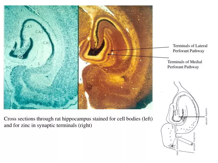
Terminals of Lateral Perforant Pathway Terminals of Medial Perforant Pathway Cross sections through rat hippocampus stained for cell bodies (left) and for zinc in synaptic terminals (right)
The main input to the dentate gyrus and CA3 of the hippocampus from the cortex is called the perforant path. It consists of two distinct fiber systems from the medial and lateral entorhinal cortex, which terminate in the middle and outer thirds of the dendritic field respectively. The two sets of terminals stain differently for zinc. They differ markedly in their resting levels of release probability (p), and consequently in their transmitter release dynamics during and following repetitive activation. When probed with pairs of stimuli in the interval range of 20 msec – 4 sec, the relative degree of facilitation and depression differs markedly between the two types of terminals. This indicates that different synaptic classes may have differing resting values of p During repeated stimulation, synapses with high p run down quickly, whereas low p synapses are better able to maintain a constant response
E.C.: For a given class of synapse, synaptic depression leads to a frequency dependent steady state of transmitter release, regardless of initial conditions. This effect results in a frequency-independent steady state depolarization at the postsynaptic cell. This has an important implication for neural signaling because it means that during prolonged periods of presynaptic activity the postsynaptic cell cannot “read” the input frequency. Differences in input frequency can only be ‘read’ over about 100-200 msec. Changes in input frequency can lead to complex postsynaptic responses
Functional consequences of synaptic depression: Synaptic depression limits the duration of 'sharp-wave' bursts in hippocampus Raw EEG 7 Hz 150 Hz Spikes CA1 Population Discharge -1 0 1 seconds Behavior Rest Rate of spontaneous EPSCs is related to p x n At the end of a sharp-wave burst, spontaneous EPSC frequency is very low, indicating synaptic depression. Rate increases with time constant characteristic of the recovery of n in paired pulse studies
The first evidence that LTP involves an associative process. It was found that converging presynaptic inputs cooperated to create sufficient postsynaptic depolarization to induce LTP. When stimulated in a correlated fashion (stripes), two separate synaptic input pathways (medial and lateral) produced more reliable and more robust LTP than either pathway stimulated separately (dots). Increasing stimulus strength increases the recruitment of converging inputs. Below a threshold stimulus strength, LTP could not be induced. Therefore converging inputs cooperate.
Synapse specificity of LTP is crucial for the storage of information. Early experimental studies established both presynaptic and postsynaptic specificity in both pyramidal cells and dentate gyrus granule cells. Presynaptic specificity means only those synapses from a given axon that were coactive with other inputs to the same postsynaptic neuron undergo LTP. Postsynaptic specificity means that only coactive synapses on the same postsynaptic neuron undergo LTP. The same specificity applies to LTD.. Recent studies suggest that these specificity conditions may sometimes be violated (e.g., in developing brain), but overall they appear to be maintained at most synapses
Long-Term Depression of hippocampal synapses (LTD): If synapses could only be strengthened, this might lead to a state of hyper-ability. Recovery from LTP could be a gradual spontaneous process (“forgetting”) or it may involve an active process of competition. Some forms of learning may explicitly involve competition for synaptic strength.
Induction of LTD in adult hippocampus with a brief pairing is input-specific and NMDA receptor-dependent. a, Synaptic stimulation was paired to 30 mV in one pathway (filled symbols). Superimposed traces are averages of 10 successive evoked (paired-pulse stimulation) EPSCs in paired (left) and unpaired (right) pathways recorded in a representative cell before (thin trace) and 60 min after (thick trace) pairing. b, Summary graph of five whole-cell recordings in which synaptic stimulation was paired to 30 mV in one pathway in the presence and after washing out of 200 the NMDA receptor antagonist AP-5 (filled symbols). Superimposed traces are averages of 10 successive evoked (paired-pulse stimulation) EPSCs in the paired pathway recorded in a representative cell as indicated in the graph. Trace 1 is thin and trace 2 is thick in 1,2; trace 2 is thin and trace 3 is thick in 2,3. Calibration bars: 20 msec, 50 pA.
'Metaplasticity' Figure 7. The voltage-response function for the induction of LTD and LTP varies with the initial state of the synapse. Superimposed voltage-response curves for the induction of LTD and LTP in potentiated, naive, and depressed synapses. The putative part of the curve in potentiated synapses is shown with stripped lines. From depressed to potentiated synapses, and + slide away from each other (arrows), progressively opening the voltage window for LTD induction.
late-phase long-term potentiation (L-LTP) involves intracellular Ca2+ release. A: 4 trains of 100 Hz/1 s tetanization (arrows) induced L-LTP that was significantly reduced when intracellular Ca2+ release was blocked by perfusion with either thapsigargin (10 µM) or ryanodine (10 µM) for 40-50 min before and 30-40 min after the tetanization (black bar). Insets: representative field EPSPs before and 3 h after the tetanic stimulation. Scale bars: 5 ms, 1 mV. B: 3-train tetanization induced L-LTP that was also significantly reduced by prolonged perfusion with ryanodine (10 µM). C: 1-train tetanization induced early-phase LTP (E-LTP) that was not attenuated by prolonged perfusion with either thapsigargin (10 µM) or ryanodine (10 µM). D: 1-train tetanization alone did not induce L-LTP. However, when ryanodine receptors were activated by perfusion with ryanodine (1 µM) for 10 min before and after the tetanus, it induced stable L-LTP. Early and Late LTP, and stabilization: LTP occurs in the presence of protein synthesis inhibitors, but it fades over about an hour. Lasting LTP requires protein synthesis and also release of intracellular Ca++. In some studies, LTP has been reversed by stimulus conditions that induce LTD, but there appears to be a window of time after which this reversal becomes more difficult The Journal of Neurophysiology Vol. 88 No. 3 September 2002, pp. 1270-1278