Download
1 / 1
10 likes | 83 Views
Phantom. Phantom Holes Diameter 1.0 cm, depth 1.25 cm (D) Diameter 0.6 cm, depth 0.6 cm (B, F). Reconstructed energy (pixel size 1.2 mm x 1.2 mm). Black boxes = target and control voxels. Reconstructed energy (pixel size 2.4 mm x 2.4 mm). Radiography Studies for Proton CT.

E N D
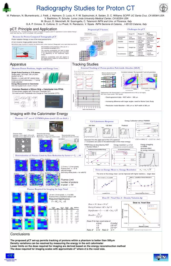
Phantom Phantom Holes Diameter 1.0 cm, depth 1.25 cm (D) Diameter 0.6 cm, depth 0.6 cm (B, F) Reconstructed energy (pixel size 1.2 mm x 1.2 mm). Black boxes = target and control voxels Reconstructed energy (pixel size 2.4 mm x 2.4 mm) Radiography Studies for Proton CT M. Petterson, N. Blumenkrantz, J. Feldt, J. Heimann, D. Lucia, H. F.-W. Sadrozinski, A. Seiden, D. C. Williams SCIPP, UC Santa Cruz, CA 95064 USAV. Bashkirov, R. Schulte Loma Linda University Medical Center, CA 92354 USAM. Bruzzi, D. Menichelli, M. Scaringella, C. Talamonti INFN and Univ. of Florence, Italy G.A. P. Cirrone, G. Cuttone, D. Lo Presti, N. Randazzo, V. Sipala INFN Sezione di Catania, , I-95123 Catania, Italy pCT: Principle and Application R. W. Schulte, et al.,, “Conceptual design of a proton computed tomography system for applications in proton radiation therapy”, IEEE Trans. Nucl. Sci., vol51, no.3, pp 866 – 875, June 2004. Challenges for pCT Proposed pCT System Reasons for Proton Computed Tomography pCT Proton radiation therapy is one of the most precise forms of non-invasive image-guided cancer therapy. Well defined range of protons in material, low entrance dose, a dose maximum (“Bragg peak”) and a rapid distal dose fall-off, One limitation to the precision: is the use of x-ray computed tomography (CT). The resulting uncertainties can lead to range errors from several millimeters up to more than 1 cm depending on the anatomical region treated. Additional uncertainties exist with respect to the target position relative to normal tissues in the treatment room. Apparatus Tracking Studies External Tracking of Proton predicts Path inside Absorber (MLP) Measure Proton Positions, Angles and Energy Loss The most likely path of an energetic charged particle through a uniform medium D C Williams Phys. Med. Biol. 49 (2004) 2899–2911 Single ProtonTracking in 10 Si planes: Single-sided, 192 strips, 236 mm pitch, [GLAST 97 B.T.) Module = x-y pair with 90o rotated strips Entrance and exit telescopes + 1 “roving” inside absorber Energy Measurement in Calorimeter One CsI crystal 5 cm x 5 cm x 15 cm Correlation between “Roving plane# 2” and exit parameters Measure displacement with “Roving Plane”at 3 depth s s s s 200 200 and 2 and 2 path’ path’ Common Readout of Silicon Strip + Calorimeter into FPGA Si have binary readout with Time-over-Threshold (ToT) CsI read out with Photodiode into Charge-to-Time-Converter (CTC) Displacement in the “Roving planes” vs. exit displacement bins of 500 mm (all angles). MLP (size of open symbol = MLP spread). • Good agreement data – MLP within ~ 300 mm • Increasing difference with larger angles: need for Monte Carlo Study • Resolution inside Absorber < 500 mm vs. MLP width of 380 mm Imaging with the Calorimeter Energy Phantom = 6th out of 12 PMMA plates (each 1.25 cm thick ) Energy Calibration without PMMMA CsI Calorimeter Response 200 MeV 100 MeV Pedestal Gaussian fit to the upper part of the CTC spectrum ( ¼ of protons are useful in the determination of the mean energy) Energy straggling In PMMA dominant effect for energy resolution. PMMA Data not described by NIST -> Nuclear Effects ? Energy resolution E in CsI (measured and corrected for pedestal) Determination of Fluence Limit by Data Reduction by factor n = 2,…, 64 (1.2 mm x 1.2 mm pixels) Fluence limit : n = 8-16 (blurred image and many white pixels = no valid fit) Fluence Limit number of protons in pixel > 10! Error on Energy Mean vs. Energy Resolution The error on the energy mean can be improved with higher statistics = larger dose Fluence Required for Imaging the large Voxel ( 8 mm x 8 mm pixels, n =1 to 200) Fluence limit is reached at about n=64 Required Significance >2 Dose D – Voxel Size d – Density Variation Dr Dose D for two voxel sizes d: d [cm] D [mGy] 1.2 2.8*10-5 0.6 2.5*10-4 Ratio d3 0.13 Ratio D 0.11 Dr = 1.2 g/cm3. “counting limit”: requirement of at least 10 protons in the fit Conclusions The proposed pCT set-up permits tracking of protons within a phantom to better than 500mm Density variations can be resolved by measuring the energy in the exit calorimeter Lower limits on the dose required for imaging are derived based on the energy reconstruction method The dose required for imaging scales with approximate d-3 where d is the voxel size.