Download
1 / 48
500 likes | 847 Views
Respiratory System Diseases. Pathophysiology. Review of Anatomy & Physiology. Respiratory Mucosa lined with ciliated mucus producing cells 125cc/ day purifies air is contiguous with all structures Nose paranasal sinuses frontal, maxillary, sphenoid, ethmoid lighten skull

E N D
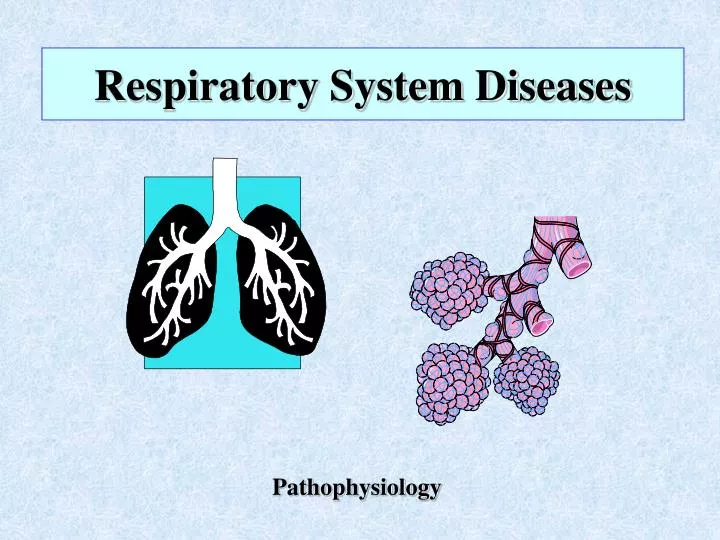
Respiratory System Diseases Pathophysiology
Respiratory Mucosa lined with ciliated mucus producing cells 125cc/ day purifies air is contiguous with all structures Nose paranasal sinuses frontal, maxillary, sphenoid, ethmoid lighten skull sound resonant chambers conchae (3 pairs) warm & humidify air lacrimal ducts olfactory receptors Pharynx 3 parts: Nasopharynx, Oropharynx, Laryngopharynx Tonsils(3 pairs) pharyngeal (adenoids) palatine lingual Eustachian (auditory) tubes open into nasopharynx equalizes pressure between middle ear & the outside Larynx composed of pieces of cartilage Thyroid cartilage= Adam’s apple epiglottis & glottis Upper Respiratory Tract
Lower Respiratory Tract • Trachea • composed of C- shaped cartilaginous rings • called windpipe • Bronchi, Bronchioles, Alveolar Duct, Alveoli • gas exchange occurs in alveoli • occurs via Passive Diffusion • Respiratory Membrane • 2 cell layers thick • surfactant = reduces surface tension to keep alveoli distended • lining of alveolus (alveolar epithelium) • lining of capillary ( capillary endothelium)
Lungs & Pleura • right lung = 3 lobes; left lung = 2 lobes • lower part of lung resting on diaphragm = Base of lung • upper part of lung under clavicle = Apex of lung • pleura = serous membrane (i.e. secretes some fluid) • parietal pleura lines thoracic cavity • visceral pleura lines organs (viscera) • Mechanics of Breathing • air moves by differences in air pressure • Inspiration • active process; get contraction of diaphragm & external intercostal muscles • results in increase in size of chest cavity • Expiration • passive process with normal expiration • active process with forced expiration; get contraction of abdominal & internal intercostal muscles • results in decrease in size of chest cavity which increases pressure & forces air out
Manifestations of PulmonaryDisease • Sneezing = reflex response to irritation of upper respiratory tract • Coughing = reflex response to irritation of lower respiratory tract • Sputum production • If yellowish- green ------ infection • If rusty ------- blood + pus = pneumococcal pneumonia • If bloody , called “hemoptysis” ---- usually frothy --- seen in pulm. Edema • Also seen in pulm. TB & cancer • Large amounts & foul = bronchiectasis • Thick & sticky = asthma, cystic fibrosis • Breathing patterns • Eupnia, labored (dyspnea) , wheezing, stridor • Breath sounds • Normal, rales, rhonchi, decreased breath sounds • Dyspnea --- discomfort feeling when can’t get enough air • Orthopnea = dyspnea lying down • Cyanosis --- not a reliable early indicator of hypoxia
Infectious diseases Upper URI Croup Epiglottitis Flu (Influenza) Lower Bronchiolitis (RSV) Pneumonia SARS TB Fungal diseases Obstructive lung diseases Cystic fibrosis Cancer Aspiration pneumonia Asthma COPD (chronic obstr. pulm. dis) Emphysema Chronic bronchitis Restrictive lung diseases Chest wall abnormalities Connective tissue abnormalities Pneumoconioses Vascular disorders Pulmonary edema Pulmonary embolism Expansion disorders Atelectasis Pleural effusion Pneumothorax Resp. distress syndrome Infant adult Respiratory System DiseasesGeneral Outline
Upper Respiratory Tract Infections Upper Respiratory Infection (URI) • Def = Acute inflammatory process that affects mucus membrane of the upper respiratory tract • Includes one or more of the following problems • Rhinitis ------ also called Coryza • Pharyngitis • Laryngitis • Sinusitis • Pathophysiology ------ see next slide • Sx = low-grade fever, malaise, cephalgia, sore throat, & discharge • Incubation period short ----- 2-3 days • As a rule: bacterial diseases = short incubation viral diseases = long incubation except URI’s • Etiol = over 200 different viruses have been implicated • can get secondary bacterial infection • Tx = symptomatic
Influenza • 3 viral types---- A,B, &C • They mutate constantly thus preventing effective immune defense for prolonged time periods • short incubation ---- 3 days • distinguishing features from simple URI • high fever in flu (usually lasts 4-5 days) • flu gives both an upper & a lower resp. tract infections at the same time, whereas URI just gives upper tract infection • Death may result from pneumonia
3 Respiratory Infections of Children • Laryngotracheobronchitis (croup) ----- upper tract infection • Neonate to 3 years • Virus --- usually adenovirus • Begins as URI; then get inspirational stridor & “barking” cough • Self-limited; last 3 days • Epiglottitis ---------- upper tract infection • 3 – 7 years • Bacterial; H. influenzae • Get drooling and dysphagia • Med emergency • Bronchiolitis (respiratory syncytial virus [RSV] ) ---- lower tract infection • 2 mon - 12 mon (young infants) • Virus --- RSV myxovirus • Get necrosis & inflammation in small bronchi & bronchioles • Get cough, wheezing, and dyspnea • If partial obstruction of bronchioles ----- get “air trapping” • If complete obstruction of bronchioles ---- get atelectasis • Tx = supportive (see next slide)
RSV Infection --- bronchiolitis • Approx. 50% of all children admitted for lower resp. tract infection have RSV • By age three, 100% of American kids infected • Predisposing factors: • Family hx of asthma • Cigarette smoke • Treatment: • Usually supportive • Have antivirals one can use (ribavirin) • In late 1990’s got updated RSV immune globulin • Indicated for prophylaxis against RSV • Monoclonal antibody to RSV • Indications: high risk kids (prematurity, bronchopulmonary dysplasia, etc) • Cost = $900 • Clinically: • Can get air trapping (hyper-inflation) • Can get obstructive atelectasis
Lower Respiratory Tract Infections Pneumonia • 6th leading cause of death in US; incidence highest in elderly • etiol • pathogens ------ bacteria, mycoplasma, viruses, fungi • Note: most all cases are preceded by URI • Common bacteria: pneumococcus • Viral = commonest in children • Community acquired: • Pnenmococcus, mycoplasma, Hi-b • Nosocomial • Pseudomonas , staph aureus (MRSA) • trauma to lungs • FB aspiration • poison • Types • Bronchopneumonia – diffuse • Lobar pneumonia – pneumococcus --- get consolidation & pleurisy • Primary atypical pneumonia (PAP) --- interstitial ---- viral & mycoplasma (see next slide)
Pneumonia (cont) • Some different types • Legionnaires Disease • etiol = gram negative bacteria (Legionella) • thrives in warm moist aquatic environment • * cooling towers of air conditioning systems • *hot water plumbing of buildings • spread by inhalation • Diagnosisdifficult since hard to culture & requires special media • Tx = antibiotics • Primary atypical pneumonia (PAP) • Pathophysiology = interstitial inflammation • Etiol = mycoplasma (occasionally viral) • Diagnosis = difficult • Onset vague with nonproductive cough • Commonest = older children & young adults • Pneumocystis carinii pneumonia (PCP) • Opportunistic infection in AIDS • Microbe is a fungus • Treatment = bactrim or septra
Pneumonia (cont) • Fungal Pneumonia ------ 3 types • (1)Coccidiomycosis ------ from soil, southwest US • (2)Histoplasmosis -------- from soil, midwest • (3)Blastomycosis --------- from soil, southeast US • grow in soil where there is bird & chicken excretion • spread = airborne (inhalation of fungal spores) • Generally, primary infection is self-limiting & heals spontaneously • may get disseminated disease ---- called secondary stage • Lung granuloma with necrosis & spread to other areas • Can become quite serious • Complication of pneumonia • Pulmonary Abscess • etiol = complication of pneumonia, neoplasm, or aspiration (food) • commonest site = lower lobe right lung • Tx = ? Incision & Drainage (I&D)
Severe Acute Respiratory Syndrome (SARS) • First diagnosed in China and reported as a atypical pneumonia (Feb 2003) • Etiol = coronavirus • Transmission: droplets during close contact • Short incubation (1 week) • Type = interstitial pneumonia • Tx: ribavirin & steroids • Mortality: 10% - 50% • Dx: difficult since get no antibodies for 3 weeks (incubation = only 1 week)
Tuberculosis • TB was in declining incidence in US until late 1980’s when it’s incidence began to increase • Why? • HIV population & opportunistic infections • homeless population • increase in low socio-economic population • These three became new reservoir for the TB bacillus • doctor complacency • drug co. complacency • patient non-compliance • these last 3 led to bacterial resistance ( bacteria evolved) • Etiol = tubercle bacillus which is quite resistant to eradication and can live in a dry form(inactive) for long times --- i.e. a spore-like state • Example = dried sputum • very contagious via air droplets • Course of the Disease • Initial Infection ( called Primary TB) often asymptomatic • patients are NOT contagious at this time • Secondary TB is reactivation and may be years later • Seen when patient’s resistance is lowered • Key to diagnosis is a positive culture; skin tests = PPD, Mantoux, Tine • If + Skin test, then do chest x-ray
TB (cont) • Signs & symptoms • Primary TB = asymptomatic • Secondary TB\ • 10% of cases will become symptomatic • Key = night sweats • Develop productive cough (purulent & bloody) • Becomes increasingly severe • Inhalation • Tubercle formation • Caseation necrosis • Called: Ghon complex • Cavitation
Obstructive Lung Diseases Cystic Fibrosis • Also called mucoviscidosis • Etiol: genetic autosomal recessive • Gene on 7th chromosome • Dx: sweat test
Lung Cancer • leading cause of cancer deaths in both men & woman in US • 4 cell types • Oat Cell (2) Squammous Cell (3) AdenoCa (4) lg cell • Pathophysiology • Most arise from bronchi or bronchioles • Squamous cell from large bronchi at hilus of lung • Slow growing; late metastasis • Adenocarcinoma (bronchoalveolar) • Found in periphery • Present frequently with pleural effusion • Moderate growth rate • Oat cell (small cell) • Found centrally near large bronchus • Fast growing & early metastasis • Large cell (undifferentiated) • Rapid growth & early metastasis • Found in periphery thus get pleural effusion
Lung Cancer (cont) • Effects from lung cancer • Obstruction • Inflammation • Pleural effusion • Paraneoplastic syndrome • The tumor cells secrete hormone-like substances • ADH • ACTH • Squamous cell lung cancer
Squamous cell cancer Oat cell cancer Adenocarcinoma Large cell cancer
Aspiration Pneumonitis • Def: passage of foreign material into trachea &/or lungs • Commonest site = right lower lobe (most direct route) • Solid objects produce obstruction • Liquid objects produce inflammation • Complications: • Acute respiratory distress syndrome • Pulmonary abscess • Systemic effects of absorbed material • Etiol: • Newborn meconium • Infants & foreign bodies • Children with congenital abnormalities (e.g. cleft palate) • Adults at operation • Adults with “café coronary” --- talking while eating • Old people with loss of gag reflex
Asthma • Def: periodic episodes of reversible bronchial obstruction in people with hypersensitive airways • Path ---- 1. Bronchospasm & get constricted bronchi , wheezing • 2. Bronchial inflammation & get edema , mucorrhea • 3. Mucorrhea leads to obstructive plugs • 2 key pathogenetic mechanisms: • Bronchospasm & inflammation
Asthma (cont) • Etiology = immune system gone awry !!! • Inflammatory reaction to an antigen from allergen or irritant exposure • Then antigen- antibody reaction ; key antibody = IgE • Allergens & irritants include: • respiratory irritants such as dust & cigarette smoke • exercise, especially in cold weather • allergies • URI &other lung infections • This causes mast cells to release: • Histamines which constricts bronchi & cause vessels to leak fluid thus producing edema and congestion • Leukotrienes which combine to form SRS-A • Inflammatory cytokines • Chronic asthma can lead to irreversible damage to lung tissue from frequent attacks • Incidence of asthma has increased over last 10 – 15 years • Status asthmaticus
Chronic Obstructive Pulmonary Disease (COPD) • get irreversible progressive obstruction of air flow in lungs • Includes • (1) Chronic Bronchitis (bronchiectasis) • (2) Emphysema • (3) Chronic asthma • General Treatment Plan • (1) broncodilators (4) anti-inflammatories • (2) mucolytic agents (5) antihistamines • (3) expectorants (6) antibiotics
Chronic Bronchitis (COPD) • def = chronic inflammation of mucus membrane • this results in a chronic, deep, productive cough • path = hyperplasia of mucosa & destruction of cilia • etiol = long term smoking, certain environmental factors such as • textile dust fibers • Sx = productive cough, SOB, wheezing • Bronchiectasis (COPD) • def = permanent, irreversible dilation & distortion of bronchi • etiol • chronic irritation producing chronic bronchitis • complication of cystic fibrosis • TB • Takes years to develop ; Primarily in the lower lobes • Sx = chronic productive cough, halitosis
Emphysema (COPD) • Def = destructive disease of alveolar septa • permanent & irreversible • get dilated non-functioning alveoli • Etiol = any chronic lung condition, pollution • Sx • insidious onset, get progressive dyspnea • moist productive cough • to exhale must use accessory muscles, thus get “barrel chest” • patients frequently “purse lips” to exhale ; this causes circumoral cyanosis • Dx • chest x-ray shows: translucent appearing lungs, flattened diaphragm, & cardiomegaly • clubbed fingers • CHF (right sided) with resultant distended neck veins and hepatomegaly
Restrictive lung disorders • 2 groups of diseases • Abnormalities of chest wall which limits lung expansion • Includes: • Kyphosis • Scoliosis • Polio • ALS • Muscular dystrophy • Disease affecting lung tissue that provides supporting framework • Includes: • Occupational diseases (pneumoconioses) • Idiopathic pulmonary fibrosis (autoimmune disease) • Pulmonary edema • Acute respiratory distress syndrome (ARDS)
Pneumoconiosis • Def = occupational diseases from inhaling inorganic dust particles over a long time period (10 years or greater) • Pathophysiology • Get inflammation & fibrosis • This destroys the connective tissue framework of lungs • Lung compliance is lost • Of all the causes, asbestosis is worst • Also gives one pleural fibrosis & lung cancer • Sx : • First to appear is dyspnea on exertion • Eventually get pulmonary hypertension • Types • Anthracosis = black lung disease from coal dust (carbon) • Asbestosis = from asbestos fibers, most common form • Berylliosis = from beryllium dust (semiconductors) • Silicosis = from silica dust (quartz dust) e.g. stone quarries Asbestos bodies in lung
Pulmonary edema Pathophysiology Fluid collection (edema) in all lung tissues Affects gas exchange Affects lung expansion Key = pulmonary capillary pressure increases & fluid moves into alveoli Capillaries rupture & get bloody sputum (hemoptysis) True medical emergency Etiology Left sided heart failure Hypoproteinemia Inhalation of toxic gases Lymphatic blockage (e.g. from tumor) Vascular disorders
Pulmonary Emboli • def = clot of foreign matter that occludes artery in pulmonary system • Size of embolus & general health of patient determine degree of damage and amount of symptoms • see next slide for pathophysiology • etiol • determined by composition of emboli • thrombus (most common) , air, fat, bacteria, tissue • risk increased by CHF, lung disease, stasis with varicosities • 90% originate from deep veins (primarily in leg) • Old age large bone fractures give fat emboli • Sx • generally apprehension, cough, chest pain, fever • if severe ------ dyspnea, tachypnea, hemoptysis • if massive ----- shock & death • Dx = imaging, blood gases • Tx • maintain adequate ventilation via O2 & anticoagulants • ? Thrombolytic drugs • Prevention via early ambulation, TED Stockings
Expansion disorders • Atelectasis • not a disease, but a condition where you get collapsed pulmonary tissue • this condition results in degrees of hypoxia • Sx • dyspnea • if large area involved get anxiety, diaphoresis, substernal retraction, cyanosis • Etiol see next slide • (1) obstruction of bronchial tree via, CA, FB, inflammation • Obstruction leads to absorption atelectasis • (2) pleural effusion • (3) inadequate effect at breathing • in newborn via prematurity or maternal narcosis • Post-op • (4) compression atelectasis • Tx = IPPB, suction & O2 if obstruction
Pleural effusion & pleuritis • Pleurisy (Pleuritis) • def = inflammation of membranes that surround lungs and pleural cavity---i.e. parietal & visceral pleura • etiol = secondary to other diseases, esp. pneumonia & cancer • can also be secondary to trauma • 2 types • Wet Type ------- get increase fluid in space & thus lung • compression with pain on inspiration • Dry Type -------- get decrease fluid, thus get friction • when breathing (friction rub) • Sx = pain with inspiration & shallow, rapid breaths
Pleural Effusion • “wet” pleuritis results in pleural effusion • Pathophysiology = fluid separates the two pleural membranes and thus the lungs do not expand properly during inspiration since no cohesion between ---- lung/visceral pleura/ parietal pleura • Large effusions give mediastinal shift • Large effusions increase pressure in mediastinum & impair cardiac filling • Types: • Transudates = hydrothorax • Blood = hemothorax • Pus = empyema
Pneumothorax • def = collection of air or gas in pleural cavity resulting in collapse ( either partial or full) of lung • Etiol & types • Spontaneous pneumothorax • from blebs, • from too much pressure with ventilation, • from tumor • Open pneumothorax --- usually secondary to trauma • Tension pneumothorax • Both of the above may result in this serious typo • Sx = sudden pain & shock-like symptoms • Tx = thoracentesis with chest tube see next slides
Flail Chest • def = double fracture of 3 or more adjacent ribs • Sx = segment involved moves in with inspiration & out with expiration, thus paradoxical movement of ribs • etiol = trauma, auto steering wheel • Temporary treatment = stabilize flail section of thorax with heavy object to prevent outward flaring
RDS (Respiratory Distress Syndrome) • 2 Types: • Infant RDS(also called: hyaline membrane disease) • leading cause of death in premies • etiol = not enough surfactant • First appears in early 3rd trimester & is adequate by 37 weeks • Amount determined by L/S ratio • lecithin-sphingomyelin • Done via amniocentesis • Treatment = synthetic surfactant (Exosurf Neonatal)
2 Types: • Adult RDS (currently called: Acute Respiratory Distress Syndrome(ARDS) • Called “shock lung” • def = alveoli fill with exudate & get respiratory failure • etiol = inhalation of things that ruin surfactant & inflame endothelium • exp = water, smoke, vomit, chem fumes • pathophysiology = alveolar & capillary wall injury • Inflammatory reaction to noxious agents that affects: • Type II alveolar cells (they produce surfactant) • Capillary endothelial cells