Download
1 / 47
470 likes | 821 Views
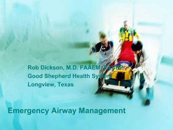
Emergency Airway Management Pat Melanson, MD. Safe airway management. airway evaluation identification of the difficult airway assessment of other clinical factors selection of the likely most successful plan of action reasonable alternative plan. Algorithmic Approach to Airway Management.

E N D
Safe airway management • airway evaluation • identification of the difficult airway • assessment of other clinical factors • selection of the likely most successful plan of action • reasonable alternative plan
Algorithmic Approach to Airway Management • Have a precompiled plan of airway management ready for implementation as clinical airway difficulties are encountered • develop a plan and a back-up plan • Practice guidelines for management of the difficult airway • ASA taskforce • Anesthesiology 78 : 597 - 602, 1993
Emergency Airway • full stomach • altered level of consciousness • deteriorating cardiorespiratory physiology • abnormal or distorted upper airway anatomy • no time for pre-assessment or plan
Airway Assessment • compromise or threats • potentially difficult airway
The Three Pillars of Airway Management • Patency ( airflow integrity ) • Protection against aspiration • Assurance of oxygenation and ventilation
Indications for Active Airway Intervention • Patency - relief of obstruction • Protection from aspiration • Hypoxic/ hypercapnic respiratory failure • Airway access for pulmonary toilet, drug delivery,therapeutic hyperventilation • Shock
Clinical Signs of Airway Compromise : Patency • Inspiratory stridor • Snoring ( pharyngeal obstruction ) • Gurgling ( foreign matter/ secretions ) • Drooling ( epiglottitis ) • Hoarseness ( laryngeal edema/ vc paralysis) • Paradoxical chest wall movement • Tracheal tug
Clinical Signs of Airway Compromise : Protection • Blood in upper airway • Pus in upper airway • persistant vomiting • Loss of protective airway reflexes
Clinical Signs of Airway Compromise:Oxygenation and Ventilation • Central cyanosis • Obtundation and diaphoresis • rapid shallow respirations • Accessory muscle use • Retractions • Abdominal paradox
The Difficult Airway • Difficult laryngoscopy • Difficult bag-mask ventilation • Lower airway difficulty
Techniques for the Compromised Airway • Bag-Valve-Mask Ventilation • Endotracheal Intubation • Rapid Sequence Intubation • Alternate techniques for the difficult airway
Golden Rules of Bagging • “ Anybody ( almost ) can be oxygenated and ventilated with a bag and a mask “ • The art of bagging should be mastered before the art of intubation • Manual ventilation skill with proper equipment is a fundamental premise of advanced airway management
Frequent Errors with BVM • failure to recognize its importance • forget to bag ( focussed on ETT ) • give up on bagging too early • bag but don’t assess efficacy • failure to assign one person to airway management only
Difficult Airway : BVM • Upper airway obstruction • Lack of dentures • Beard • Midfacial smash • facial burns, dressings, scarring • poor lung mechanics
Difficult Airway : BVM • degree of difficulty from zero to infinite • zero = no external effort/internal device • one person jaw thrust/ face seal • oropharyngeal or nasopharyngeal AW • two person jaw thrust / face seal • both internal airway devices • infinite -no patency despite maximal external effort and full use of OP/NP
Difficult Airway : BVM • Remove FB - Magill forceps • Triple maneuver if c-spine clear • Head tilt, jaw lift, mouth opening • Nasopharyngeal or oropharyngeal airway • two-person, four-hand technique
Prediction of the difficult airway (Intubation) • 1200 prospectively studied patients • of 84 patients predicted to have problem, only 22 (25%) actually had a problem • of 43 actual difficult intubations incurred, only 22 (51%) were predicted • Latto IP. and Rosen M
Prediction of the difficult airway • history of past airway problems • Careful physical assessment • knowledge and experience to overcome the "unpredicted difficult airway". • learning practical airway management skills in an environment that is not urgent, stressful or life threatening
Difficult Airway : Laryngoscopy • Short thick neck • Receding mandible • Buck teeth • Poor mandibular mobility/ limited jaw opening • Limited head and neck movement • ( including trauma )
Difficult Airway : Laryngoscopy • Tumor, abscess or hematoma • Burns • Angioneurotic edema • Blunt or penetrating trauma • Rheumatoid arthritis, ankylosing spondylitis • Congenital syndromes • Neck surgery or radiation
Difficult Airway : Laryngoscopy • 3 fingerbreadths mentum to hyoid • 3 fb chin to thyroid notch • 3 fb upper to lower incisors • Head extension and neck flexion • Mallimpadi classification • Previous history of difficult intubation
Mallimpadi Classification ( Tongue to Pharyngeal Size ) • I - soft palate, uvula, tonsillar pillars • 99 % have grade I laryngoscopic view • II - soft palate, uvula • III - soft palate, base of uvula • IV - soft palate not visible • 100% grade III or grade IV views
Unsuccessful Intubation • Bag the patient • Maximize neck flexion/ head extension • Move tongue out of line of site • Maximize mouth opening • Look for landmarks and adjust blade • BURP maneuver • increasing lifting force • consider Miller blade • Bag the patient
Dilemmas: • Awake or Asleep • Oral or Nasal • Laryngoscopy or Blind Intubation • To Paralyze or Not
Case #1 • 43 year old female, day 12 post SAH • 5 unclipped cerebral aneurysms • vasospasm with left hemiparesis • hydrocephalus with clotted IV drain • rising ICP and BP • decreasing LOC • ate breakfast
Techniques • DL without pharmacologic aids • Awake Direct Laryngoscopy • Awake Blind Nasal • Rapid Sequence Intubation (RSI) • Fiberoptic • Surgical Cricothyroidotomy
Anesthesia Airway Maxims • the awake airway is the safest to manage • spontaneous breathing is generally safer than paralysis with PPV by mask • have a low threshold to wake the patient up and cancel the case • call for help early
The “Intubation Reflex “ • Catecholamine release in response to laryngeal manipulation • Tachycardia, hypertension, raised ICP • Attenuated by beta-blockers, fentanyl • ICP rise possibly attenuated by lidocaine • Midazolam and thiopental have no effect
Rapid Sequence Intubation :Definition • The near simultaneous administration of a sedative-hypnotic agent and a neuromuscular blocker in the presence of continuous cricoid pressure to facilitate endotracheal intubation and minimize risk of aspiration • modifications are made depending upon the clinical scenario
Rapid Sequence Intubation :Advantages • Optimizes intubating conditions/ facilitates visualization • Increased rate of successful intubation • Decreased time to intubation • Decreased risk of aspiration • Attenuation of hemodynamic and ICP changes
Rapid Sequence Intubation :Contraindications • Anticipated difficulty with endotracheal intubation • anatomic distortion • Lack of operator skill or familiarity • inability to preoxygenate
Rapid Sequence Intubation :Procedure • Pre-intubation assessment • Pre-oxygenate • Prepare ( for the worst ) • Premedicate • Paralyze • Pressure on cricoid • Place the tube • Post intubation assessment
Pre-oxygenate ( Time - 5 Minutes) • 100 % oxygen for 5 minutes • 4 conscious deep breaths of 100 % O2 • Fill FRC with reservoir of 100 % O2 • Allows 3 to 5 minutes of apnea • Essential to allow avoidance of bagging • If necessary bag with cricoid pressure
Preparation ( Time - 5 Minutes ) • ETT, stylet, blades, suction, BVM • Cardiac monitor, pulse oximeter, ETCO2 • One ( preferably two ) iv lines • Drugs • Difficult airway kit including cric kit • Patient positioning
Pre-treatment/ Prime ( Time - 2 Minutes ) • Lidocaine 1.5 mg/kg iv • Defasciculating dose of non-depolarizing NMB • Beta-blocker or fentanyl • Induction agent • Thiopental 3 - 5 mg/kg • Midazolam 0.1 - 0.4mg/kg • Ketamine 1.5 - 2.0 mg/kg • Fentanyl 2 - 30 mcg/kg
Paralyze ( Time Zero ) • Succinylcholine 1.5 mg/kg iv • Allow 45 - 60 seconds for complete muscle relaxation • Alternatives • Vecuromium 0.1 - 0.2 mg/kg • Rocuronium o.6 - 1.2 mg/kg
Pressure • Sellick maneuver • initiate upon loss of consciousness • continue until ETT balloon inflation • release if active vomiting
Place the Tube ( Time Zero + 45 Secs ) • Wait for optimal paralysis • Confirm tube placement with ETCO2
Post-intubation Hypotension • Loss of sympathetic drive • Myocardial infarction • Tension pneumothorax • Auto-peep
Succinylcholine : Contraindications • Hyperkalemia - renal failure • Active neuromuscular disease with functional denervation ( 6 days to 6 months) • Extensive burns or crush injuries • Malignant hyperthermia • Pseudocholinesterase deficiency • Organophosphate poisoning
Succinylcholine : Complications • Inability to secure airway • Increased vagal tone ( second dose ) • Histamine release ( rare ) • Increased ICP/ IOP/ intragastric pressure • Myalgias • Hyperkalemia with burns, NM disease • malignant hyperthermia
Difficult Airway Kit • Multiple blades and ETTs • ETT guides ( stylets, bougé, light wand) • Emergency nonsurgical ventilation ( LMA, combitube, TTJV ) • Emergency surgical airway access ( cricothyroidotomy kit, cricotomes ) • ETT placement verification • Fiberoptic and retrograde intubation
Emergency Surgical Airway Maxims • they are usually a bloody mess, but ... • a bloody surgical airway is better than an arrested patient with a nice looking neck
Case # 2 • 42 year old female • right Pancoast tumor • RUL, RML, RLL collapse • ARDS on left • hypoxemic respiratory failure • cord compression C7 - T4