Download
1 / 80
811 likes | 1.15k Views
Endocrine Review. Eric L. Johnson, MD Assistant Professor Department of Family and Community Medicine UNDSMHS Assistant Medical Director Altru Diabetes Center Grand Forks, ND. Objectives. Understand basic principles of endocrine function

E N D
Endocrine Review Eric L. Johnson, MD Assistant Professor Department of Family and Community Medicine UNDSMHS Assistant Medical Director Altru Diabetes Center Grand Forks, ND
Objectives • Understand basic principles of endocrine function • Understand basic principles of endocrine dysfunction • Understand management and referral of common endocrine diseases
Endocrine Pathology • Disease state results from excess or insufficiency of hormone • Clinical challenge is determination of the origin of excess or insufficiency, i.e., Hypothalamus (tertiary), Pituitary (secondary) or target gland (primary)
Endocrine Disorders (other than diabetes) • Thyroid • Adrenal • Parathyroid • Pituitary gland • Gonadal • Gout (uric acid)
The Endocrine System Brain Hypothalamus Pituitary Hypothyroid Thyroid Pancreas Adrenal Gonads
Hypothalamus AVP = arginine vasopressin CRH = corticotropin-releasing hormone GHRH = growth hormone-releasing hormone GnRH = gonadotropin-releasing hormone SRIF = somatotropin release–inhibiting factor (somatostatin) TRH = thyrotropin-releasing hormone; VIP = vasoactive intestinal polypeptide.DA = dopamine ACTH = adrenocorticotropic hormone LH = lutenizing hormone FSH = follicle-stimulating hormone GH = growth hormone TSH = thyroid-stimulating hormone PRL = prolactin Goldman: Cecil Medicine, 23rd ed. 2007
Anterior Pituitary Hormones • TSH, ACTH, FSH, and LH hormones are tropic hormones that simulate other endocrine glands • TSH-Thyroid • ACTH- Adrenal Cortex • FSH, LH- Gonads
Posterior Pituitary Hormones • Vasopressin(ADH)- kidney, baroreceptors (plasma osmolality, water retention, thirst) • Oxytocin- breast, uterus (no known function in males) • Both are synthesized in specialized neurons in the hypothalamus (neurohypophysial neurons)
Prolactinomas • Pituitary adenomas may present with visual impairment, headache, or hormonal abnormalities • Prolactinomas most common. Manifest with galactorrhea and gonadal dysfunction • Laboratory testing: serum prolactin, creatinine levels and thyroid function tests • MRI is the imaging modality of choice for the anatomic evaluation of the hypothalamus and pituitary gland
Panhypopituiarism • Neoplasm • Radiation • Infiltrative/Infection • Empty sella syndrome (herniation of subarachnoid tissue) • Apoplexy (hemorrage) • Sheehan’s syndrome (pregnancy)
Growth Hormone Excess (Acromegaly) • Acromegaly cause: growth hormone–secreting adenoma of the anterior pituitary • Elevated serum levels of IGF-1 are found in acromegaly (best single test) • The diagnosis of acromegaly is confirmed by a glucose tolerance test • Transsphenoidal surgery to remove the pituitary adenoma is the initial treatment of choice in individuals with acromegaly
Growth Hormone Excess (Acromegaly) Acromegaly manifestations • Increased hand/foot size • Prognathism/teeth space widening • Frontal bossing/coarsening of facial features • Weakness/fatigue
Growth Hormone Excess (Acromegaly) Acromegaly manifestations cont’d • Sweating • HTN>>>cardiomyopathy • Obstructive sleep apnea • Insulin resistance
Growth Hormone Excess (Acromegaly) Treatment • Surgical resection Best if tumor small, 10-20% result in pan hypo pit • Radiation Slow to diminish GH, 50% pan hypo pit • Bromocriptine Blocks GH effect • Octreotide Lowers GH secretion, SC route w/frequent dosing
Growth Hormone Deficiency • Fatigue or hypoglycemia in the adult • Dwarfism in child Born normal length at birth but growth “falls off curve” • Diagnosis: IGF1 level (GH varies too much) • Treatment: formerly used human pit gland extract, synthetic GH since late 80’s
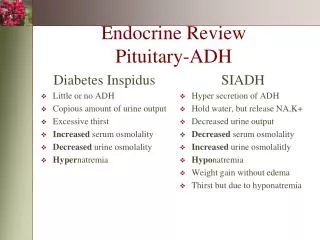
Diabetes Insipidus (ADH) • Deficiency of posterior pituitary hormone ADH (aka vasopressin) • ADH acts on kidney collecting duct to retain free water deficiency causes free water loss • ADH maintains blood volume via: - -Osmoreceptors in brain -Stretch receptors in heart -Baroreceptors in carotids and aorta
Diabetes Insipidus (ADH) • Decreased urinary specific gravity (≤1.005) • Decreased urinary osmolarity (<200 mOsm/kg) even in the presence of high serum osmolality • Hypernatremia, increased plasma osmolarity, hypercalcemia, hypokalemia • Normal Serum osmolarity: 282 - 295 mOsm/kg • Normal Urine osmolarity: 500 - 800 mOsm
Diabetes Insipidus (ADH) Etiology -CNS insult -head trauma, surgery, tumor, infection -Genetic -MS, Metastatic Disease -Drugs (lithium is classic) -Nephrogenic vs. Neurogenic vs. Psychogenic Sx –Thirst, polyuria, polydipsia (with a normal glucose)
Diabetes Insipidus (ADH) • Diagnostic workup: decreased ADH or insensitivity to ADH? • Water Deprivation Test: -Baseline measurement of weight, ADH, plasma sodium, urine and plasma osmolarity -Patient is deprived of fluids under strict medical supervision. -Frequent (q2h) monitoring of plasma and urine osmolarity • Test terminated when plasma osmolarity >295 mOsm/kg or the patient loses ≥3.5% of initial body weight. • Diabetes insipidus is confirmed if the plasma osmolarity is >295 mOsm/kg and the urine osmolarity is <500 mOsm/kg (typical referral point)
Diabetes Insipidus (ADH) • Nephrogenic vs. neurogenic • Patient given 5 U of vasopressin (ADH), change in urine osmolarity is measured • Significant increase (>50%) in urine osmolarity after administration of ADH is indicative of neurogenic diabetes insipidus.
Diabetes Insipidus (ADH) Treatment • Desmopressin • Thiazides in mild neurogenic
Syndrome of Inappropriate ADH(SIADH) • Hyponatremia • Urinary osmolarity > serum osmolarity • Normal BUN, creatinine,TSH,glucose
SIADH • Neoplasm • Pulmonary disorders: pneumonia, emphysema, cystic fibrosis, status asthmaticus, respiratory failure • Intracranial pathology: trauma, neoplasms, infections (meningitis, encephalitis, brain abscess) • Postoperative period: surgical stress, ventilators with positive pressure, anesthetic agents • Drugs: chlorpropamide, thiazide diuretics, chemotherapeutic agents carbamazepine, phenothiazines, MAO inhibitors, tricyclic antidepressants, narcotics, nicotine, clofibrate, haloperidol, SSRIs, NSAIDs • Other: acute intermittent porphyria, myxedema, psychosis, delirium tremens, ACTH deficiency (hypopituitarism), general anesthesia, endurance exercise
SIADH • Treatment Fluid Restriction Careful use of hypertonic saline IV Resolution/Treatment of underlying problem
Cushing’s Disease • Pituitary adenoma>>excess production of ACTH>>excess cortisol production • Distinguished from Cushing’s syndrome, which includes other causes of cortisol excess (ectopic production of ACTH and CRH) • Cushing’s disease causes 60-70% of excess cortisol disease states • Occurs 8 times more often in women than men
Cushing’s • Classic Cushing's features: Centripetal obesity, moon facies, and ‘buffalo’ hump • Striae are common • Fine (lanugo) hair growth • Muscle wasting • Bone demineralization • Hypertension • IGT • Psych
Cushing’s Diagnosis • Distinguish between: • Cushings Disease -Pituitary Causes • Cushing’s Syndrome -Adrenal causes of cortisol excess; -Ectopic sources of ACTH or Ectopic CRH (Cortisol Releasing Hormone)
Cushing’s Diagnosis • Serum Cortisol is elevated • Abnormal tests can be seen in up to 30% of hospitalized and/or depressed patients • 24 hour free urinary cortisol can be a useful adjunct • Overnight Dexamethasone suppression test……
Cushing’s Diagnosis • Overnight Dexamethasone Suppression Test: 1 gram at 11pm, measure plasma cortisol at 8 am the next morning • The normal response is suppression to less than 3mcg/dl • If no suppression, they have ectopic or adrenal production • If supression, they may have pituitary cause
Cushing’s Diagnosis • Can Measure ACTH -ACTH low in Adrenal gland tumor -ACTH high in ectopic or pituitary adenoma
Cushing’s DiagnosisSummary • High serum or 24 hour urine cortisol • Dexamethasone suppression -No supression: Ectopic or adrenal -Supression: Pituitary • ACTH -Low: Adrenal -High: Likely pituitary or ectopic
Addison’s Disease(Adrenocortical Hypofunction) • Can result in all loss of corticosteroid production if the adrenal cortex suffers destruction (primary) • Can result from diminished ACTH production (secondary)
Addison’s Disease • Loss of cortisol: -Loss of vascular tone and CV output -Hypoglycemia-Cortisol important for Gluconeogenesis -Hypercalcemia (Loss of inhibition of intestinal absorption and renal reabsorption) -Serum ACTH levels are usually used for initial screening (Low)
Addison’s Disease • Skin changes-Hyperpigmentation in Palmar creases, scars, oral mucosa • Longitudinal pigmented bands under nails • Vitiligo in up to 15% of patients • Decreased pubic and axillary hair in females • Weakness, fatigue, nausea and vomiting, and a craving for salt
Addison’s Disease • Associated with other endocrine insufficiencies (thyroid, parathyroid, type 1 DM, etc) • Treatment is to replace adrenal hormones • Case I’ve seen: Pt. also had Type 1 DM and hypothyroidism; died at age 27 from profound hypoglycemia
Aldosterone Disorders • Aldosterone-a mineralocorticoid secreted by the adrenal glands • Primary secretion affected by Angiotensin II>renin (part of fluid and electrolyte balance) • Increased aldosterone-increased sodium retention and increased potassium secretion by the kidney
Hyperaldosteronism • Primary-more common in women 3rd to 5th decade of life • Presents with hypertension, weakness, fatigue, hypokalemia, polyuria, polydipsia • Most cases are from benign adenomas (Conn’s Syndrome) • Screening: aldosterone:renin ratio of greater than 30 (off of anti-hypertensives, except Ca++ channel blockers) • CT scanning for adrenal adenomas
Secondary Hyperaldosteronism • Usually occurs in edematous states-i.e. CHF, cirrhosis or renal artery stenosis • Causes intravascular volume depletion, stimulating renin production • Elevated renin and aldosterone levels • Can occur in Bartter’s syndrome (impaired chloride re-absorption)
Hypoaldosteronism • Sodium wasting and hyperkalemia • May be up to 10% of hyperkalemia • Hyper-reninemic hypoaldosteronism (more common) Defect is in aldosterone synthesis or angiotensin II action Genetic, ACEI, ARB, heparin, Lead poisoning, Severe Illness • Hyporeninemic DM, HTN, renal insufficiency
Hypoaldosteronism • Lab: Plasma renin K+ Glucose Kidney functions Hyperchloremic metabolic acidosis
Parathyroid • Purpose –Maintain serum calcium levels • Target tissues –Bone, kidney, intestine • Feedback loop –As Calcium rises, PTH lowers –As calcium lowers, PTH rises
Hyperparathyroidism • Etiology –Tumor (adenoma) –Hyperplasia –Drugs (lithium is classic) –Ectopic PTH • Secondary Hyperparathryroidism can occur in chronic renal disease
Hyperparathyroidism • Elevated PTH, Serum Ca++, urine Ca++ • Polyruia, Polydipsia • Kidney stones • Peptic ulcer disease • Pancreatitis • Nausea, vomiting or loss of appetite • Osteopenia/porosis, leading to an increased risk of fractures • Confusion or poor memory • Muscle weakness or fatigue
Hyperparathyroidism • Treatment (loop diuretics, hydration) • Observation • Surgery
Hypoparathyroidism • Low PTH (idiopathic, iatrogenic-thyroid surgery) • Low Serum Ca++ (low Vit D?) • Elevated Phosphorous • Parasthesthias • Alopecia/ vitiligo/ candidiasis • Long Q-T on EKG • Muscle cramps or tetany -Chvostek’s sign: facial twitch after a gentle tapping over the facial nerve -Trousseau's sign: carpopedal spasm after inflation of blood pressure cuff above the patient's systolic blood pressure for 2 to 3 minutes
Hypoparathyroidism • Ca++ plus Vitamin D • Low Phosphorous diet
Hypothyroidism • Incidence in the U.S. is about 1% • Primary hypothyroidism accounts for 90-95% of all cases • Autoimmune most common (Hashimoto’s) • May or may not have enlarged (goitrous) thyroid • End Stage Grave’s/treatment/can result in hypothyroidism • Iatrogenic-surgical • Iodine deficiency
Hypothyroidism • Decreased secretion of thyroid hormone from the thyroid gland. Most frequently reflects a disease of the gland itself (primaryhypothyroidism) 95% • Pituitary disease (secondaryhypothyroidism) • Hypothalamic disease (tertiaryhypothyroidism)