Download
1 / 19
220 likes | 656 Views
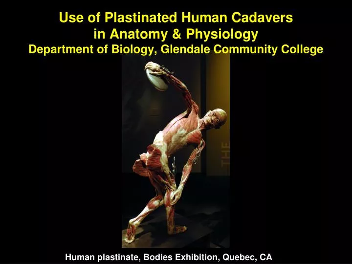
Use of Plastinated Human Cadavers in Anatomy & Physiology Department of Biology, Glendale Community College. Human plastinate, Bodies Exhibition, Quebec, CA. What is a human plastinate?. A human plastinate is a cadaver that has been preserved with a dry, odorless plastic polymer

E N D
Use of Plastinated Human Cadaversin Anatomy & PhysiologyDepartment of Biology, Glendale Community College Human plastinate, Bodies Exhibition, Quebec, CA
What is a human plastinate? • A human plastinate is a cadaver that has been preserved with a dry, odorless plastic polymer • Method developed by Gunther von Hagen (German anatomist) • Can study real human anatomy without the drawbacks of conventional formaldehyde-preserved specimens
Disadvantages of Traditional Methods • Odor • Cost • Lack of student participation • Need elaborate facility with ventilation William and Mary Sports Medicine Program Anatomy Lab Pennsylvania College of Technology Physician’s Assistant Program Anatomy Lab
Advantages of Plastinates • Lack of odor • More student participation • Last indefinitely (so low cost) • Using real specimens is motivational - students find real specimens inherently more interesting than plastic models. • Using real specimens shows natural variation – models represent an average and collections of models are identical, but real specimens demonstrate the significant variation seen among individuals. Institute for Plastination, Heidelberg Germany
Plastinated specimens can be used for teaching students about human organs and organ systems. Even minute details and textures are preserved as in the delicately folded lining of the intestines. Institute for Plastination, Heidelberg Germany
The New GCC Life Science Building The Anatomy & Physiology labs were designed for the use of plastinated specimens.
The Plastination Process • Tissue preservation technique invented by Gunter von Hagen in 1985: • Remove water and fatty tissues with a series of solvents • Remove solvents under vacuum and replace with a liquid plastic • Cure plastic with a catalyst • Specimen will be flexible if silicone is used, or hard if an expoxy or polyester resin is used • Specimens are dry, odorless and last indefinitely • Can be handled and stored like a plastic model http://www.koerperspende.de/en/plastination/
Sources of Plastinated Specimens • The UofAPlastination Laboratory is a high quality facility that produces plastinated human specimens for users inside and outside the University. • Equipped to do both standard silicone impregnation and epoxy sheet plastination. • The Body Donor Program can provide specimens for users to dissect and return to the laboratory for finishing if they cannot produce find their own specimens. • Administered by Joshua Lopez, LFP Plastination LaboratoryDepartment of Cellular and Molecular Medicine University of Arizona College of Medicine
Corcoran Laboratories, located in Traverse City, Michigan has worked with the University of Michigan to make plastinated cadaver specimens available to colleges and universities. Plastination LaboratoryOffice of Medical Education University of Michigan Medical School The UM Plastination Laboratory is a cost-for-service unit that produces human plastinate specimens for users inside and outside the University; Administered by Dr. Ameed Raoof
The collection of plastinates in the Biology Department at GCC currently includes a torso, upper and lower limbs, hearts, brains, knees, intestines, liver, pancreas, kidneys, tongue and larynx.The following photos of GCC plastinates were taken by Dr. Rob Bowker NOTE: The following slides show plastinated human body parts and may be graphic in their nature. Viewer discretion is advised.