Download
1 / 35
520 likes | 1.64k Views
Arthrogryposis and Amyoplasia. Mohammed T. Attiah, MD November 10 th - 2003. Definition. Arthrogryposis Group of unrelated diseases with the common phenotypic characteristic of multiple congenital joint contractures Amyoplasia “Symmetric contractures” = AMC IR shoulder

E N D
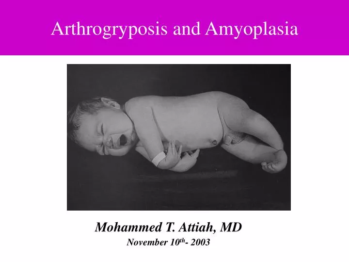
Arthrogryposis and Amyoplasia Mohammed T. Attiah, MD November 10th- 2003
Definition • Arthrogryposis • Group of unrelated diseases with the common phenotypic characteristic of multiple congenital joint contractures • Amyoplasia “Symmetric contractures” = AMC • IR shoulder • Extended elbow, flexed hand and wrist • Knee “extended or flexed” • Talipes equinovarus • Dislocated hips…. Stern WG: Arthrogryposis multiplex congenita. JAMA 1923
Epidemiology & Etiology • AG- 1:3,000 AP- 1:10,000 • Arthrogryposis is multifactorial etiology: • Fetal akinesia,Curare Injection.Drachman DB, Lancet 1962 • Viral infection, alkaloid ingestion • Hyperthermia, Oligohydromanios, AHC defect, Myopathy • Amyoplasia is sporadic ??“Genetic” • Larsen’s syndrome • Distal arthrogryposis type I & II
Differential Diagnosis • Full H & P and limbs-spine x-ray • Amyoplasia is relatively easy to recognize • Spine x-ray “spinal dysraphism” • CPK “Congenital M Dystrophy” • CT brain “Structural brain anomalies” • Chromosomal studies, experienced geneticist • Muscle biopsy and EMG ??? myopathy
Amyoplasia • Four limbs 84% • Lower limbs 11% • Upper limbs 5% Sells JM,. Pediatrics 1996 • Joint have limited ROM, firm,and inelastic end point • Trunk generally spared, although scoliosis 30% Sarwark JF, J bone Joint Surg Am 1990
Amyoplasia • Muscle mass • Fusiform limbs • Lack of normal skin creases over the joint • Webbing across elbow & knees • Skin dimpling on the extensor muscle • Sensation N, DTR diminished or absent • Midline facial hemangioma and micrognathia • Inguinal hernia, cryptochridism • Abdominal wall defect, Gastrochisis, Bowel atresia
General Management • Overall function is related to • Family support • Patient personality • Education early efforts to foster independence Carlson WO,. Clin Orthop 1985 • Parents “Walking” • Helps parents focus on factors that will substantially improve the child’s function • Upper extremities Vs Lower extremities
General Management • Gentle stretching and ROM exercise • Lightweight splinting “ acceptable joint position “ • Casting or ST release and casting • Muscle transfer • Nonfunctioning muscles ?? • Functioning muscles “ limited excursion “ • Osteotomy • Skeletal maturity “Recurrence of the deformity “
Upper Extremity Deformities • Provide an extremity that can be brought to the mouth and stabilized for feeding and to provide for toilet care or pulling up from sitting position Williams PF, Clin Orthop 1985 • Where is the problem: • Shoulder IR ? osteotomy • Lack of active elbow flexion ± elbow extension contractures
Non-Surgical Treatment • Passive stretching is most successful to obtain motion • Shoulder, wrist and fingers are the most resistant • Elbow stretching • Mild change in ROM will substantially improve the ability to • Dress • Self-feed • Personal hygiene • Passive elbow flexion “TRICKS “
Surgical Management of the Upper extremity Deformities • Defer most surgery until the patient is old enough to demonstrate functional achievement Lloyd-Roberts GC,,, J Bone Joint 1970
Elbow Contractures • Elbow flexion < 90° with supervised elbow stretching • Posterior capsulotomy with triceps lengthening • Post-op passive elbow flexion maintained for two years • Intra-articular incongruity ??? Van Heest A, J Hand Surg 1998
Tendon Transfer Indications • Age > 4 • Lack of active flexion • Minimum of 90° passive elbow flexion • Ipsilateral hand motion • Absent contralateral active elbow flexion • Available donor muscle • Triceps-to-biceps transfer gives most reliable results Van Heest A,. J Hand Surg 1998 Contraindication: Ambulate or transfer in lower limbs involved child Complication: Elbow flexion contractures Carroll RE, JBJS 1970
Elbow Contractures • P. Major transfer • Best donor in the absence of triceps • Large surgical scar “ sternum to anticubital fossa” • Breast asymmetry Schottstaedt ER, J Bone Joint Surg 1955 • Steindler Flexorplasty • Flexor tendon are weak Doyle JR,. J Hand Surgery 1980
Wrist Deformities • Early release and casting for wrist flexion contractures • Wrist extensor are absent • FCU only functioning muscle • FCU transfer will give wrist extension • Passive ROM “neutral” • Quengel cast hinge • PRC and tendon transfer • Wrist fusion
Feet Deformities • Rigid clubfeet • Aggressive ST release “ not lengthening “ before walking • Complete correction intra-op • Long-term bracing, night bracing, AFO • Recurrence rate 70% Niki H, J Pediatr Orthop 1997
Relapsed Clubfoot • Talectomy • Primary procedures in severe cases • Tibiocalcaneal incongruity • Loss medial column • Failed CC fusion-------- Midfoot Adduction • Reduce ST -------Foot dorsiflexion Green ADL, J Bone Joint Surg [Br] 1984
Relapsed Clubfoot • Verebelyi-Ogston procedure “ Talus Decancellation” • Maintain medial column • Avoid progressive midfoot adduction • Easier triple Spires TD, J Pediatr Orthop 1984
Relapsed Clubfoot • Circular-Frame Fixator • Tech. Demanding, good results • Trans-epiphyseal pin locked to the tibial frame “ Epi. separation” • Incision parallel to the direction of distraction Brunner R, J Pediatr Orthop B 1997
Knee Deformities • Most difficult • FC > EC • 50% FC pt = community walker • 10% EC pt = community walker Murray C, J Pediatr Orthop B 1997
Treatment of Knee Flexion Contractures • Stretching • Bracing • Casting “ ? posterior tibia dislocation” • Quengel hinge • Point of rotation • Tibia move forward with extension
Treatment of Flexion Contractures “Surgical” • Posterior ST release ± shortening osteotomy • Muscles planes “ fibrous dens cord “ • No tornique “ facilitate vascular dissection” • II incision PM & PL, avoid S-incision • Anterior release • PF adhesion “Rug under the door” • Medial patellar incision • Gradual correction • Full correction….. ??NV structure • Hyperextension = Hypertension
Recurrent Knee contractures • Supracondylar extension osteotomy ± shortening • Immediate correction • Dog leg-type deformity • Cosmetically unacceptable • Recurrence 1°/month in Sk immature patients DelBello DA, J Pediatr Orthop 1996
Knee Extension Contractures • Walk well • Sitting difficulty • Difficulty rising from a chair • Treatment: • Quads percutaneous release + casting • Quadricepslasty + Knee open reduction
Hip Deformities • Hip problems in arthrogryposis 65-80% • Flexion contractures common, dislocation 15-30% Sarwark JF, J Bone Joint Surg Am 1990 • Hip FC ----Lumbar lordosis • ER contractures “Do not correct” = gait stability • Hip FC > 45° ---- surgical release
Hip Dislocation • Teratologic • Poor results with CR • Options • Acceptance of dislocation • Open reduction “ medial or anterior “ • well-performed open reduction • Redislocation, stiffness, and AVN Szoke G, J Pediatr Orthop 1996 Cruel CR, J Pedaitr Orthop 1986
Twenty-Years F/U of Hip Problems in Arthrogryposis Multiplex Congenita, Peter W.P. Yau, JPO 2002, Hong Kong • Unilateral hip dislocation • Openly reduced hips are stiffer • 121° Vs 103° • Long term hip function score was comparable • 69 Vs 73; P= 0.174
Hip Dislocation • Unilateral dislocation should perform open reduction 6-12 • Best results with medial approach Szoke G, J Pediatr Orthop 1996 Cruel CR, J Pedaitr Orthop 1986 • Bilateral dislocation ?????? • Supple hip that is dislocated is preferable to a reduced but stiff hip
Spine Deformities • Scoliosis 30-67% • Poor prognosis for progression: • Early curve onset • Paralytic curve pattern • Pelvic obliquity • Quiet stiff curve • Posterior fusion = 35% correction • Post + Ant. = 44% correction • Pseudo-arthrosis 15- 30% Yingsakmongkol W, J Pediatr Orthop 2000
Arthrogryposis • Hips & Foot deformities • Early and aggressive with surgical treatment
Arthrogryposis • Knee deformities • Be cautious with surgical treatment
Arthrogryposis • Upper extremity deformities • Be very careful with the surgical treatment