Download
1 / 46
1.25k likes | 2.98k Views
Common Lower Limb Deformities in Children. Prof. Mamoun Kremli AlMaarefa College. Objectives. Angular deformities of LLs Bow legs Knock knees Rotational deformities of LLs In-toeing Ex-toeing Feet problems. Angular LL Deformities of LL . Nomenclature. Bow legs. Knock knees.

E N D
Common Lower Limb Deformities in Children Prof. Mamoun Kremli AlMaarefa College
Objectives • Angular deformities of LLs • Bow legs • Knock knees • Rotational deformities of LLs • In-toeing • Ex-toeing • Feet problems
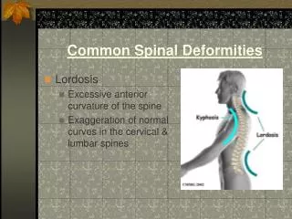
Nomenclature Bow legs Knock knees Genu Varus Genu Valgus
Normal range varies with age • During first year: Lateral bowing of Tibiae • During second year: Bow legs (knees & tibiae) • Between 3 – 4 years: Knock knees
Evaluation Should differentiate between • “physiologic” and “pathologic” deformities
Evaluation Physiologic Pathologic • Symmetrical • Asymmetrical • Mild – moderate • Severe • Progressive • Regressive • Generalized • Localized • Expected for age • Not expected for age
Physiologic Pathologic Causes • Normal for age • Rickets • Exaggerated : • Endocrine disturbance • Metabolic disease - Overweight • Injury to Epiphys. Plate • - Infection / Trauma - Early wt. bearing - Use of walker? • Idiopathic
Evaluation Symmetrical deformity
Evaluation Asymmetrical deformity
Evaluation Generalized deformity
Evaluation Localized deformity Blount’s
Evaluation Localized deformity Rickets Improves in time
Assess angulation - standing/supine Bow Legs (genu varus) • Inter-condylar distance
Assess angulation - standing/supine knock knees (genu valgus) • Inter-malleolar distance
Measure angulation - standing/supine Use Goniometer • Measure angles directly • More accurate • More appropriate
Investigations / Laboratory • Serum Calcium / Phosphorous ? • Serum Alkaline Phosphatase • Serum Creatinine / Urea – Renal function
Investigations / Radiological • X-ray when severe or possibly pathologic • Standing AP film: • long film (hips to ankles) with patellae directed forwards • Look for diseases: • Rickets / Tibia vara (Blount’s) / Epiphyseal injury.. • Measure angles
Investigations / Radiological Medial Physeal Slope Femoral-Tibial Axis
When To Refer ? • Pathologic deformities: • Asymmetrical • Localized • Progressive • Not expected for age • Exaggerated physiologic deformities • Definition ?
Rotational LL Deformities In-toeing / Ex-toeing • Frequently seen • Concerns parents • Frequently prompts varieties of treatment • often un-necessary / incorrect
Rotational Deformities • Level of affection: • Femur • Tibia • Foot
Femur • Ante-version = more medial rotation • Retro-version = more lateral rotation
Normal Development • Femur: Ante-version: • 30 degrees at birth • 10 degrees at maturity • Tibia: Lateral rotation: • 5 degrees at birth • 15 degrees at maturity
Normal Development • Both Femur and Tibia laterally rotate with growth in children • Medial Tibial torsion and Femoral ante-version improve ( reduce ) with time • Lateral Tibial torsion usually worsens with growth
Clinical Examination • Rotational Profile • At which level is the rotational deformity? • How severe is the rotational deformity? • Four components: • Foot propagation angle • Assess femoral rotational arc • Assess tibial rotational arc • Foot assessment
Rotational Profile • Foot propagation angle – Walking • Normal Range: ( +10o to -10o ) • ? In Eastern Societies • Normal range: ( +25o to - 5o ) Fundamentals of Pediatric Orthopedics, L Stahili
Rotational Profile • Assess femoral rotation arc Supine Extended
Rotational Profile • Assess femoral rotation arc Supine Flexed
Rotational Profile • Assess tibial rotational arc • Foot-thigh angle in prone
Rotational Profile • Foot assessment • Metatarsus adductus • Searching big toe • Everted foot • Flat foot
Common Presentations • Infants: out-toeing • Toddlers: In-toeing • Early childhood: In-toing • Late childhood: Out-toing
Infants: out-toeing • Normal • seen when infant positioned upright • (usually hips laterally rotate in-utero) • Metatarsus adductus: • medial deviation of forefoot • 90% resolve spontaneously • casting if rigid or persists late in 1st year Fundamentals of Pediatric Orthopedics, L Stahili
Toddlers: In-toeing • Most common during second year • (at beginning of walking) • Causes: • Medial tibial torsion: does not need treatment • Metatarsus adductus: if sever, casting works • Abducted great toe: resolves spontaneously
Rotational DeformitiesCommon PresentationsChild • In-toeing : due to medial femoral torsion • Out-toeing : in late childhood • lateral femoral / tibial torsion
Medial Femoral Torsion • Starts at 3 - 5 years • Peaks at 4 – 6 years • Resolves spontaneously by 8-9years • Girls > boys • Look at relatives - family history – normal • Treatment usually not recommended • If persists > 8 years and severe, may need surgery
Medial Femoral Torsion (Ante-version) • Stands with knees medially rotated • (kissing patellae) • Sits in “W” position • Runs awkwardly (egg-beater) Family History
Lateral Tibial Torsion • Usually worsens • May be associated with knee pain (patellar) • specially if LTT is associated with MFT • (knee medially rotated and ankle laterally rotated) Fundamentals of Pediatric Orthopedics, L Stahili
Medial Tibial Torsion • Less common than LTT in older child • May need surgery if : • persists > 8 year, • and causes functional disability Fundamentals of Pediatric Orthopedics, L Stahili
Management of Rotational Deformities • Challenge : dealing effectively with family • In-toeing: • Spontaneously corrects in vast majority of children as LL externally rotates with growth • Best Wait !
Management of Rotational Deformities • Convince family that only observation is appropriate • Only < 1 % of femoral & tibial torsional deformities fail to resolve and may require surgery in late childhood
Management of Rotational Deformities • Attempts to control child’s walking, sitting and sleeping positions is impossible and ineffective, cause frustration and conflicts • Shoe wedges and inserts: • ineffective • Bracing with twisters: • ineffective - and limits activity • Night splints: • better tolerated - ? Benefit
Management of Rotational Deformities Shoe wedges Ineffective Twister cables Ineffective Fundamentals of Pediatric Orthopedics, L Stahili
When To Refer ? • Severe & persistent deformity • Age > 8-10y • Causing a functional disability • Progressive
Summary • Angular deformities are common: • Genu varus • Genu valgus • Differentiate between physiologic and pathologic deformities • Rotational deformities are common • Part of normal development • In-toing Vs Out-toing • Cause may be in femur, tibia, or foot • Most improve with time