Download
1 / 1
10 likes | 227 Views
With and Without the Cell Cytoskeleton. Intermediate Filaments

E N D
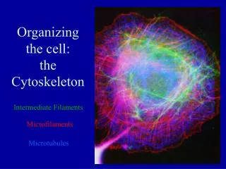
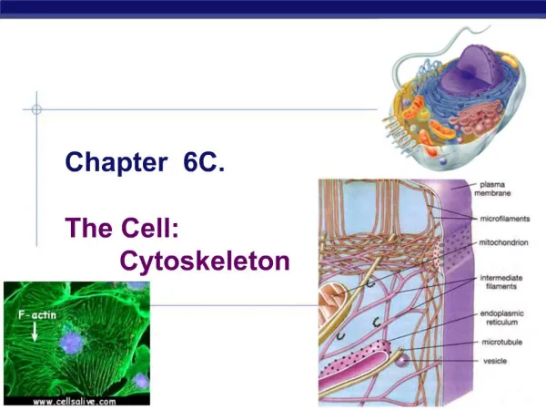
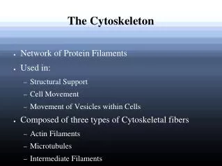
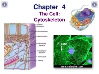
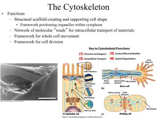
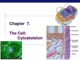
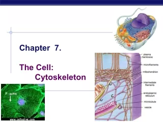
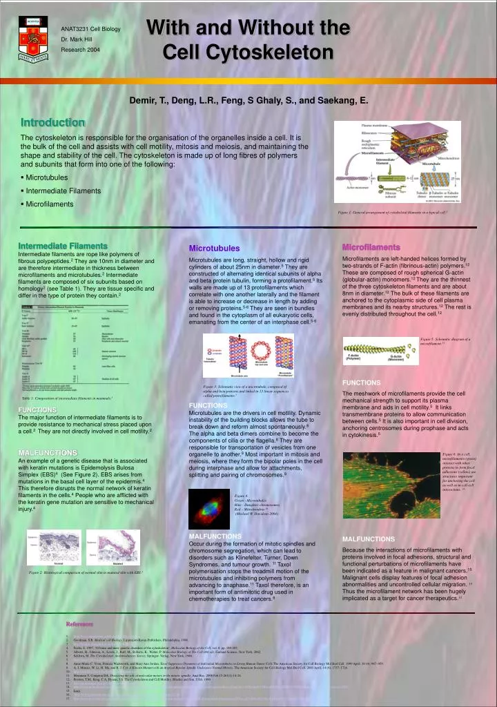
Withand Without the Cell Cytoskeleton Intermediate Filaments Intermediate filaments are rope like polymers of fibrous polypeptides.2 They are 10nm in diameter and are therefore intermediate in thickness between microfilaments and microtubules.2 Intermediate filaments are composed of six subunits based on homology2 (see Table 1). They are tissue specific and differ in the type of protein they contain.2 FUNCTIONS The major function of intermediate filaments is to provide resistance to mechanical stress placed upon a cell.2 They are not directly involved in cell motility.2 MALFUNCTIONS An example of a genetic disease that is associated with keratin mutations is Epidemolysis Bulosa Simplex (EBS)4 (See Figure 2). EBS arises from mutations in the basal cell layer of the epidermis.4 This therefore disrupts the normal network of keratin filaments in the cells.4 People who are afflicted with the keratin gene mutation are sensitive to mechanical injury.4 Table 1: Composition of intermediate filaments in mammals.3 Figure 2: Histological comparison of normal skin to mutated skin with EBS.3 ANAT3231 Cell Biology Dr. Mark Hill Research 2004 Demir, T., Deng, L.R., Feng, S Ghaly, S., and Saekang, E. • Introduction • The cytoskeleton is responsible for the organisation of the organelles inside a cell. It is the bulk of the cell and assists with cell motility, mitosis and meiosis, and maintaining the shape and stability of the cell. The cytoskeleton is made up of long fibres of polymers and subunits that form into one of the following: • Microtubules • Intermediate Filaments • Microfilaments Figure 1: General arrangement of cytoskeletal filaments in a typical cell.1 Microfilaments Microfilaments are left-handed helices formed by two-strands of F-actin (fibrinous-actin) polymers.12 These are composed of rough spherical G-actin (globular-actin) monomers.12 They are the thinnest of the three cytoskeleton filaments and are about 8nm in diameter.12 The bulk of these filaments are anchored to the cytoplasmic side of cell plasma membranes and its nearby structures.12 The rest is evenly distributed throughout the cell.12 FUNCTIONS The meshwork of microfilaments provide the cell mechanical strength to support its plasma membrane and aids in cell motility.5 It links transmembrane proteins to allow communication between cells.5 It is also important in cell division, anchoring centrosomes during prophase and acts in cytokinesis.5 MALFUNCTIONS Because the interactions of microfilaments with proteins involved in focal adhesions, structural and functional perturbations of microfilaments have been indicated as a feature in malignant cancers.15 Malignant cells display features of focal adhesion abnormalities and uncontrolled cellular migration. 15 Thus the microfilament network has been hugely implicated as a target for cancer therapeutics.15 Microtubules Microtubules are long, straight, hollow and rigid cylinders of about 25nm in diameter.5 They are constructed of alternating identical subunits of alpha and beta protein tubulin, forming a protofilament.5 Its walls are made up of 13 protofilaments which correlate with one another laterally and the filament is able to increase or decrease in length by adding or removing proteins.5-6 They are seen in bundles and found in the cytoplasm of all eukaryotic cells, emanating from the center of an interphase cell.5-6 FUNCTIONS Microtubules are the drivers in cell motility. Dynamic instability of the building blocks allows the tube to break down and reform almost spontaneously.8 The alpha and beta dimers combine to become the components of cilia or the flagella.8 They are responsible for transportation of vesicles from one organelle to another.9 Most important in mitosis and meiosis, where they form the bipolar poles in the cell during interphase and allow for attachments, splitting and pairing of chromosomes.9 MALFUNCTIONS Occur during the formation of mitotic spindles and chromosome segregation, which can lead to disorders such as Klinefelter, Turner, Down Syndromes, and tumour growth. 11 Taxol polymerisation stops the treadmill motion of the microtubules and inhibiting polymers from advancing to anaphase.11 Taxol therefore, is an important form of antimitotic drug used in chemotherapies to treat cancers.8 Figure 5: Schematic diagram of a microfilament.13 Figure 3: Schematic view of a microtubule, composed of alpha and beta proteins and linked in 13 linear sequences called protofilaments.7 Figure 6: In a cell, microfilaments (green) interact with other proteins to form focal adhesions (yellow) are structures important for anchoring the cell as well as in cell-cell interactions. 14 Figure 4: Green - Microtubules Blue - Daughter chromosomes Red – Mitochondria 10 (Michael W. Davidson 2004) • References • http://bio.winona.msus.edu/bates/Bio241/cells.htm • Goodman, S.R. Medical cell Biology. Lippincott-Raven Publishers, Philadelphia, 1998. • http://hykim.chungbuk.ac.kr/lectures/biomem/19/19-1.htm • Fuchs, E. 1997, ‘Of mice and men: genetic disorders of the cytoskeleton’, Molecular Biology of the Cell, vol. 8, pp. 189-203. • Alberts, B., Johnson, A., Lewis, J., Raff, M,, Roberts, K., Walter, P. Molecular Biology of The Cell (4th ed). Garland Science, New York, 2002. • Schliwa, M. The Cytoskeleton: An Introductory Survey. Springer- Verlag, New York, 1986. • http://wine1.sb.fsu.edu/BCH4053/Lecture30/Lecture30.htm • Anne-Marie C. Yvon, Patricia Wadsworth, and Mary Ann Jordan. Taxol Suppresses Dynamics of Individual Microtubules in Living Human Tumor Cells.The American Society for Cell Biology Mol Biol Cell. 1999 April; 10 (4): 947–959. • A. I. Marcus, W. Li, H. Ma, and R. J. Cyr. A Kinesin Mutant with an Atypical Bipolar Spindle Undergoes Normal Mitosis. The American Society for Cell Biology Mol Biol Cell. 2003 April; 14 (4): 1717–1726. • http://146.201.224.61/cells/fluorescencemitosis/ • Mountain V, Compton DA. Dissecting the role of molecular motors in the mitotic spindle. Anat Rec. 2000 Feb 15;261(1):14-24. • Preston, T.M., King, C.A, Hyams, J.S. The Cytoskeleton and Cell Motility, Blackie and Son, USA. 1990 • http://micro.magnet.fsu.edu/cells/plants/microfilaments.html • http://www.ncbi.nlm.nih.gov/entrez/query.fcgi?cmd=Search&db=books&doptcmdl=GenBookHL&term=intermediate+filaments+AND+mcb%5Bbook%5D+AND+107004%5Buid%5D&rid=mcb.section.5543 • Lucy • http://www.pubmedcentral.gov/articlerender.fcgi?tool=pmcentrez&artid=25218 • http://www.ncbi.nlm.nih.gov/entrez/query.fcgi?cmd=Search&db=books&doptcmdl=GenBookHL&term=microfilaments+AND+cell%5Bbook%5D+AND+8041%5Buid%5D&rid=cell.figgrp.4674