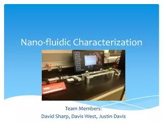
Download
1 / 23
380 likes | 968 Views
Microscopy as a Means for Nano-Characterization. By Thomas Williams Phys 3500. What is Microscopy?. Microscopy is any technique for producing visible images of structures or details too small to otherwise be seen by the human eye. What is Nano Characterization?. What does it look like?

E N D
Microscopy as a Means for Nano-Characterization By Thomas Williams Phys 3500
What is Microscopy? • Microscopy is any technique for producing visible images of structures or details too small to otherwise be seen by the human eye.
What is Nano Characterization? • What does it look like? • Dimensions, structure, • What is it made of? • Molecules, elemental proportions • What are it’s properties? • Physical, chemical, electromagnetic
Why Microscopy? • In order to effectively study something or build something it is important to see exactly what it is we’re doing. • As the things we are interested in get smaller and smaller we need more better, meaning more powerful microscopy. • Eventually this will necessitate advances in the physics.
The Origens of Microscopy • In the first century AD Romans invented glass and began experimenting with various shapes, stumbling upon the converging lens. • In approx. 1590 Dutch eyeglass makers Hans and Zacharias Jensonn makes a compound microscope. • Mid 17th century Anton Van Leeuwenhoek uses an improved single lens microscope to view and describe bacteria, protozoan, etc. http://www.cas.muohio.edu/~mbi-ws/microscopes/history.html
Age of the Optical Microscope • In the late 17th century Robert Hooke added a third lens, greatly improving contrast issues and comfort. • Over the next two hundred years optical microscopy revolutionizes science, especially biology. • During this time improvements are continually made, including corrections for chromatic spherical aberrations. • In the late 19th century, Ernst Abbe showed that the improvement of the magnification of optical microscopes was fundamentally limited by the wavelength of light. http://www.microscope-microscope.org/images/BWScope.jpg
History of Electron Microscopy • 1931- Ernst Ruska co-invents the electron microscope. • 1938- 10nm resolution reached. • 1940- 2.4 nm resolution. • 1945- 1.0nm resolution achieved. • 1981- Gerd Binning and Heinrich Rohrer invent the scanning tunneling electron microscope (STM). • 1986- The Atomic Force Microscope was developed in collaboration between IBM and Stanford University.
Transmission Electron Microscope (TEM) • Same principle as optical microscope but with electrons. • Condenser aperture stops high angle electrons, first step in improving contrast. • The objective aperture and selected area aperture are optional but can enhance contrast by blocking high angle diffracted electrons • Advantages: we can look at non conducting samples, i.e. polymers, ceramics, and biological samples. http://www.unl.edu/CMRAcfem/temoptic.htm
TEM Images http://www.abdn.ac.uk/emunit/emunit/temcells/index.htm
Scanning Electron Microscope (SEM) • The SEM functions much like an optical microscope but uses electrons instead of visible light waves. • The SEM uses a series a series of EM coils as lenses to focus and manipulate the electron beam. • Samples must be dehydrated and made conductive. • Images are back and white. http://www.mos.org/sln/SEM/works/slideshow/semmov.html
SEM Images http://www.mos.org/sln/SEM/works.html
Scanning Tunneling Electron Microscope (STM) • Basic principle is tunneling. • Tunneling current flows between tip and sample when separated by less than 100nm. • The tunneling current gives us atomic information about the surface as the tip scans. http://www.iap.tuwien.ac.at/www/surface/STM_Gallery/index.htmlx
What is tunneling? • The probability that the electron will exist outside the barrier in the vacuum is non zero. • If these leak-out waves overlap and a small bias voltage is applied between the tip and the sample, a tunneling current flows. • The magnitude of this tunneling current does not give the nuclear position directly, but is directly proportional to the electron density of the sample at a point. http://www.chembio.uoguelph.ca/educmat/chm729/STMpage/stmdet.htm
What does piezo-electric mean? • In 1880 Pierre Curie discovered that by applying a pressure to certain crystals he could induce a potential across the crystal. • The STM reverses this process. Thus, by applying a voltage across a piezoelectric crystal, it will elongate or compress. • A typical piezoelectric material used in an STM is Lead Zirconium Titanate.
http://www.iap.tuwien.ac.at/www/surface/STM_Gallery/index.htmlxhttp://www.iap.tuwien.ac.at/www/surface/STM_Gallery/index.htmlx
STM Images http://www.almaden.ibm.com/vis/stm/gallery.html
Atomic Force Microscopy (AFM) • AFM is performed by scanning a sharp tip on the end of a flexible cantilever across the sample while maintaining a small force. • Typical tip radii are on the order of 1nm to 10nm. • AFM has two modes, tapping mode and contact mode. • In scanning mode, constantcantilever deflection is maintained. • In tapping mode, the cantilever is oscillated at its resonance frequency. http://www.nanoscience.com/education/AFM.html http://www.azom.com/details.asp?ArticleID=3278
AFM Images http://www.azom.com/details.asp?ArticleID=3278 http://www.nanoscience.com/index.html
AFM Video http://www.nanoscience.com/education/gallery.html
Future / Conclusions • We still have a long way to go before we’ve exhausted the limits of electron wavelength resolution limit. • The wave length of a high energy electron is on the order of .001nm or 1.0pm, our current best resolution with an STM is only approximately .1nm. • Limiting factors include, aberations, contrast,
References • Wikipedia - http://en.wikipedia.org/wiki/Main_Page • History of the Microscope - http://www.cas.muohio.edu/~mbi-ws/microscopes/history.html • Molecular Expressions - http://microscopy.fsu.edu/primer/museum/hornyolddissecting1920.html • Dictionary.com - http://dictionary.reference.com/ • Micro-bus -http://www.microscope-microscope.org/microscope-home.html • BBC H2G2 - http://www.bbc.co.uk/dna/h2g2/ • About.com - http://about.com/ • MOS - http://www.mos.org/sln/SEM/works/slideshow/semmov.html • UNL - http://www.unl.edu/CMRAcfem/temoptic.htm • IBM - http://www.ibm.com/us/ • AZOM.com - http://www.azom.com/default.asp • Nanonscience Instruments - http://www.nanoscience.com/index.html
Special Thanks • Dr. Tapas Kar & the Fall 06 Nano-Chemistry Crew. • Google, and their amazing database of resources. • Utah State, for seeing the growing need to offer classes in nanotechnology.