Download
1 / 19
190 likes | 860 Views
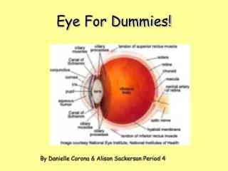
Eye For Dummies!. By Danielle Corona & Alison Sackerson Period 4 . IMPORTANT STUFF. Eyelids Eyelashes: Eyelashes are located on the top part of the eyelid help they help keep dust out of the eye.

E N D
Eye For Dummies! By Danielle Corona & Alison Sackerson Period 4
IMPORTANT STUFF Eyelids Eyelashes:Eyelashes are located on the top part of the eyelid help they help keep dust out of the eye. Conjunctiva: The conjunctiva is a membrane on the underside of the eyelid it produces tears to help keep the eye clean. Iris:The iris is the colorful part of the eye. It ahs muscles attached to is that monitor the amount of light that goes through the pupil.
IMPORTANT STUFF Continued Retina: The retina is located in the back of the eye. The retina takes the light the eye receives and changes it into nerve signals so the brain can understand what the eye is seeing. Cornea: The cornea consist of tough tissues that make up the outer layer of the eyeball and give it strength. Though the cornea has no blood vessels at all and has relatively little moisture. As a result, it is transparent The cornea enables light rays to enter the eyeball. Lens: After light goes through the pupil it hits the lens. The lens focuses light rays on the back of the eyeball.
MEDICAL ADVICE • Glaucoma is a disease in which the aqueous humor—the fluid that nourishes the cornea and the lens—does not drain properly. Pressure in the eye increases and, if untreated, destroys the optic nerve. There are two types of Glaucoma. • primary open-angle glaucoma, can cause gradual blindness and in the end total blindness. To treat glaucoma Doctors prescribe eye drops, pills, or laser therapy. Such treatments reduce pressure in the eye and so halt damage. • primary narrow-angle glaucoma or acute glaucoma can occur at any age. Symptoms include pain in the eyes or forehead and seeing things. To prevent blindness surgery or laser treatment may be necessary. .
Health Tip • Preventing eye damage. The most common injuries are caused by blows to the eye, by particles that enter the eye, and by chemical burns, explosions, and firearms. Many such injuries can be prevented. • Wear safety goggles or glasses to protect eyes during certain jobs…( construction, welding, etc.) • Wash hands before handling contacts • Do not look directly at the sun or sun lamp • Visit your optometrist (eye doctor) regularly
Interesting Facts The term color blindness is something of a misnomer. Very few (~1 in 105) people cannot distinguish colors at all. Most "color-blind" people actually have abnormal color vision such as confusing the red and green of traffic lights. As high as 8% of the males in some populations have an inherited defect in their ability to discriminate reds and greens. These defects are found almost exclusively in males because the genes that encode the red-absorbing and green-absorbing opsins are on the X chromosome. [Discussion of X-linkage
Interesting Facts The blind spot All the nerve impulses generated in the retina travel back to the brain by way of the axons in the optic nerve (above). At the point on the retina where the approximately 1 million axons converge on the optic nerve, there are no rods or cones. This spot, called the blind spot, is thus insensitive to light. Do you know how many times a day we blink? Probably not, because we blink so fast. The average person blinks about 12 times a minute. That's an amazing 10,080 blinks in a kids day (14 waking hours) http://www.optima-hyper.com/eyetests/kidsquiz/kidfact.htm
Human Body For Dummies! A successful book publisher is looking for new books to add to the “Dummies” series. Join this #1 book series by writing a “Dummies” book about the human body. The Dummies slogan is “A Reference for the Rest of Us!” Dummy books are sold by the millions because they are a fun and easy way to learn about topics that are important to everyone. This WebQuest will help you write a sample chapter for the “Human Bodies for Dummies” series. To be chosen as a writer for the new series you must demonstrate that you can take information that can be very scientific and boring and turn it into an easy and fun to read format that all will enjoy reading.
Steps to Follow 1. Select one system. Choose from: cardiovascular (circulatory) muscular skeletal the eye 2.Research facts about your system. 3.Copy notes and URL’s onto Note Cards following this page. You need to use al least 3 different websites. Add new note pages if you go to more than 3 sites. 4.Restate notes to complete your PowerPoint 5. Add pictures and final touches.
Note Card 1 URL:http://www.worldbookonline.com/wb/Article?id=ar189340&st=Human+Eye Notes:The Eyeis the organ of sight. It is our most important organ for finding out about the world around us. We use our eyes in almost everything we do-sight is our most precious sense, and many people fear blindness more than any other disability… The human eyeball measures only about 1 inch (25 millimeters) in diameter, yet the eye can see objects as far away as a star and as tiny as a grain of sand. The eye can quickly adjust its focus between a distant point and a near one. Plus, it is accurately directed toward an object even while the head is moving. Did you know the eye actually doesn’t really see objects? Instead, it absorbs the light objects reflect or give off. That’s why you can see in bright light and in dim light, but in darkness nothing is visible at all! Light rays enter the eye through transparent tissues, then the eye changes the rays into electrical signals. These signals are sent to the brain, which interprets them as visual images.
Note Card 2 Copy and Paste you research findings for each website you use. Later you will restate these notes in your project. URL:http://www.worldbookonline.com/wb/Article?id=ar189340&st=Human+Eye&sc=-1#h9 Notes:How we see For our eyes to focus, Light rays that enter the eye must come to a point on the retina for a clear visual image to form. However, the light rays that objects reflect or give off do not naturally move toward one another. Instead, they either spread out or travel almost parallel. The focusing parts of the eye—the cornea and the lens—bend the rays toward one another. The cornea provides most of the bending power of the eye. After light rays pass through the cornea, they travel through the aqueous humor and the pupil to the lens. The lens bends the rays even closer together before they go through the vitreous humor and strike the retina. Light rays from objects at which the eyes are aimed come together at the fovea centralis, a tiny pit in the center of the macula. It is the area of sharpest vision. Light rays from objects to the sides strike other areas of the retina.
Note Card 3 URL:http://users.rcn.com/jkimball.ma.ultranet/BiologyPages/V/Vision.html#blindspot Notes: In the next two cards you be amazed by the two types of visions… 1. Rod Vision- the cells that help us see movement, and at night, shades of gray. Rhodopsin is the light-absorbing pigment of the rods. It is incorporated in the membranes of disks that are neatly stacked (some 2000 of them) in the outer portion of the rod. (This arrangement is reminiscent of the organization of thylakoids, another light-absorbing device.)The outer segments of the rods contain the orderly stacks of membranes which incorporate rhodopsin. Although the disks are initially formed from the plasma membrane, they become separated from it. Thus signals generated in the disks must be transmitted by a chemical mediator (a "second messenger" called cyclic GMP (cGMP) to alter the potential of the plasma membrane of the rod. Rhodopsin consists of an opsin of 348 amino acids coupled to retinal.
Note Card 4 • URL:http://users.rcn.com/jkimball.ma.ultranet/BiologyPages/V/Vision.html#blindspot • Notes:Cone Vision • Although cones operate only in relatively bright light, they provide us with our sharpest images and enable us to see colors. Most of the 3 million cones in each retina are confined to a small region just opposite the lens called the fovea. So our sharpest and colorful images are limited to a small area of view. Because we can quickly direct our eyes to anything in view that interests us, we tend not to be aware of just how poor our peripheral vision is. • The three types of cones provide us the basis of color vision. Cones are "tuned" to different portions of the visible spectrum. • red absorbing cones; those that absorb best at the relatively long wavelengths peaking at 565 nm • green absorbing cones with a peak absorption at 535 nm • blue absorbing cones with a peak absorption at 440 nm. • Retinal is the prosthetic group for each pigment. Differences in the amino acid sequence of their opsins accounts for the differences in absorption. The response of cones is not all-or-none. Light of a given wavelength (color), say 500 nm (green), stimulates all three types of cones, but the green-absorbing cones will be stimulated most strongly. Like rods, the absorption of light does not trigger action potentials but modulates the membrane potential of the cones.
Note Card 5 • URL:http://www.worldbookonline.com/wb/Article?id=ar189340&st=Human+Eye&sc=-1#h1 • Notes: • The outer parts. The front of the eyeball is protected by the eyelids.Eyelashes on the lids block out some of the dust and other particles that might otherwise enter the eye. Any sudden movement in front of the eye—or anything that touches the eyelashes—causes the lids to blink in a protective reflex action. Also,The conjunctiva is a membrane that lines the inside of the eyelids and extends over the front of the white part of the eye. It produces mucus, a clear, slimy fluid that lubricates the eyeball. The conjunctiva also produces some tears, which help keep the eye clean. • The sclera and the cornea consist of tough tissues that make up the outer layer of the eyeball and give it strength. The sclera is the white part of the eye. Although the sclera appears to have many blood vessels on its surface, most of these vessels are part of the conjunctiva. Though the cornea has no blood vessels at all and has relatively little moisture. As a result, it is transparent. The cornea lies in front of the colored part of the eye and resembles the crystal of a wrist watch. The cornea enables light rays to enter the eyeball.
Note Card 6 • URL:http://www.worldbookonline.com/wb/Article?id=ar189340&st=Human+Eye&sc=-1#h1 • Notes: • The iris is the colored disk behind the cornea. The color of the iris comes from a brownish-black substance called melanin. • Do you know how many times a day we blink? • Probably not, because we blink so fast. The average person blinks about 12 times a minute. That's an amazing 10,080 blinks in a kids day (14 waking hours) • http://www.optima-hyper.com/eyetests/kidsquiz/kidfact.htm
Note Card 7 http://kidshealth.org/kid/body/eye_noSW.html The iris (say: eye-riss) is the colorful part of the eye. When we say a person has blue eyes, we really mean the person has blue irises! The iris has muscles attached to it that change its shape. This allows the iris to control how much light goes through the pupil (say: pyoo-pul). The pupil is the black circle in the center of the iris, and it lets light enter the eye. To see how this works, use a small flashlight to see how your eyes or a friend's eyes respond to changes in brightness. The pupils will get smaller when the light shines near them and they'll open wider when the light is gone.
Note Card 8 http://kidshealth.org/kid/body/eye_noSW.html These next parts are really cool, but you can't see them with just your own eyes! Doctors use special microscopes to look at these inner parts of the eye, such as the lens. After light enters the pupil, it hits the lens. The lens sits behind the iris and is clear and colorless. The lens' job is to focus light rays on the back of the eyeball - a part called the retina (say: reh-tin-uh). The lens works much like the lens of a movie projector at the movies. Your retina is in the very back of the eye, past the vitreous body. Though it's smaller than a dime, it holds millions of cells that are sensitive to light. The retina takes the light the eye receives and changes it into nerve signals so the brain can understand what the eye is seeing.
Publishing Resources Helpful sites to visit on the Internet for researching. World Book Encyclopedia Online The best site to start at and citations are at the ends of the articles. Medline Plus Use the Medical Encyclopedia for research. This is a layman's site produced and maintained by the National Library of Medicine (a sub group of the National Institutes on Health) that includes a dictionary, glossary, graphics, etc. on all aspects of the human body. Human Body animated anatomy At this site anatomy is animated and includes information for most systems. Human Body Systems in English and Spanish also narrated at medtroplis. Fact Monster this kid friendly site has great overviews of body systems. Web MD This site is a helpful resource for the tips and warning pages of your WebQuest it includes diseases and health topics Atlas of the Body (Pictures) http://www.ama-assn.org/ama/pub/category/7140.html