Download
1 / 43
510 likes | 930 Views
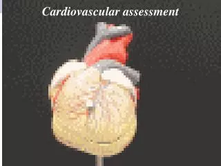
Cardiovascular Patient Assessment. J.O. Medina, RN,MSN,FNP,CCRN Education Specialist Nurse Practitioner Critical Care & Emergency Services California Hospital Medical Center. Objectives :. Outline a systematic approach to cardiovascular assessment.

E N D
Cardiovascular Patient Assessment J.O. Medina, RN,MSN,FNP,CCRN Education Specialist Nurse Practitioner Critical Care & Emergency Services California Hospital Medical Center
Objectives : • Outline a systematic approach to cardiovascular assessment. • Differentiate normal from abnormal findings when assessing the cardiovascular system. • Relate the events of the cardiac cycle to auscultatory findings.
Assessing Patient’s CV Status • History & Subjective Data • Past Medical history • Previous Illness • Diagnostic/interventional cardiac procedures • Hospitalizations • Surgeries • Allergies • AMPLE
Assessing Patient’s CV Status • CC • Common signs and symptoms of CV disease • Chest pain (most common CV symptom) • Angina • often described as “pressure” rather than pain • Usually brought by physical/emotional stress • Last: 2-5 minutes ; rarely > 20 • Relieved with rest / NTG
Assessing Patient’s CV Status • ACS (acute coronary syndrome) • Pain similar to angina ; may be more intense • Often occurs at rest • Usually last >30 minutes; usually > 2 hours • Not relieved by rest/NTG; requires analgesic • Pericarditis • May mimic ACS; often described as sharp, stabbing, shooting • Aggravated by movement • Tend to be constant • Relieved by sitting up, leaning forward, shallow breathing
Assessing Patient’s CV Status • Dyspnea • Subjective sensation of being unable to breath • Usually cause by congestion from LVF • Types: • Dyspnea on exertion (DOE) • Orthopnea : inability to breathe while lying flat • Paroxysmal nocturnal dyspnea (PND): nightime episodes of SOB due to lying flat which increases venous return (preload)
Assessing Patient’s CV Status • Fatigue / Weakness • Symptom of decreased forward CO • Usually seen as unusual fatigue at end of normal day previously tolerated • Exertional fatigue : sense of weakness or heaviness of extremities • Medications that can cause fatigue: • Diuretics : orthostatic hypotension , hypokalemia • Beta Blockers, Calcium Channel Blockers, Digoxin, antihypertensive medications
Assessing Patient’s CV Status • Fluid retention • Fluid accumulation in tissues • Common cardiac causes • Heart failure • Constrictive pericarditis • Restrictive cardiomyopathies • Weight gain of 2 lbs in 4 days or 3-5 pounds over a month may be indicative of heart failure • More severe in evening
Assessing Patient’s CV Status • Syncope/Presyncope • Temporary loss of consciousness, lightheadedness, dizziness • Cardiac cause most commonly result of inadequate cardiac output from arrythmias
Assessing Patient’s CV Status • Palpitations • Awareness of heart beat with sudden changes in rate, rhythm, increased stroke volume • Associated with : tachycardias, bradycardias, atrial fibrillation, PVCs, aortic and mitral regurgitation, signs of heart failure
Assessing Patient’s CV Status • Other symptoms • GI • Nausea, anorexia, vomiting from RVF, digoxin toxicity, inferior MI • Indigestion or flu like symptoms may be sole s/s of MI, especially in elderly or diabetic patient • Extremity pain • Intermittent claudication indicative of PVD due to decreased blood flow to muscles during time of increased demand • Ischemia from PVD
Assessing Patient’s CV Status • Other symptoms • Decreased urine output • Indicative of heart failure and hypovolemia • Look for concomitant weight gain due to CHF • Nocturia • Sign of heart failure • Caused by increased preload to heart
Assessing Patient’s CV Status • Risk Factors • Non-modifiable • Age • Sex • Family history • Race • Modifiable • Cigarette smoking • Hypertension • Hyperlipidemia • Physical inactivity • Diabetes • Stress • Obesity
endocrine function “adipokines” Leptin Pro-thrombotic Anti-inflammatory Satiety to hypothalamus Resistin Hormone making tissue insulin resistant Type II DM Adiponectin Counteracts negative effects of other hormones FAT : Adipose Tissue
Total Cholesterol < 200 mg/dL best 200 – 239 borderline high 240 mg/dL and above 2X risk of CAD Cholesterol Level : AHA Recommendation
HDL Cholesterol < 40 mg/dL (men) < 50 mg/dL (women) > 60 mg/dL cardioprotective Cholesterol Level : AHA Recommendation
LDL Cholesterol < 100 mg/dL Optimal 100 – 129 mg/dL Near or above optimal 130 – 159 mg/dL Borderline 160 – 189 mg/dL High 190 mg/dL Very high Cholesterol Level : AHA Recommendation
Triglyceride < 150 mg/dL Normal 150 – 199 mg/dL Borderline high 200 – 499mg/dL High 500 mg/dL and above Very high Cholesterol Level : AHA Recommendation
Know you’re A-B-C Numbers • Hemoglobin A1c • Measures an average BS over 3 months • Goal : under 7% • Prefer under 6.5% • Blood Pressure • < 130/80 mmHg • Cholesterol • Total : < 200 mg/dl • HDL : > 45 mg/dl in men ; 55 mg/dl in women • Triglycerides : < 150 mg/dl
Assessing Patient’s CV Status • Social History • Alcohol intake • Dietary pattern: caffeine , salt intake • Cocaine • Educational level • Medication History • Prescribed drugs • OTC
Salty Foods • Salty Foods
Physical Examination • Inspection • General appearance • Color • Cyanosis – 5 gm desaturated hemoglobin • Central Cyanosis • Decreased SaO2 – usually < 80% • Indicates cardiopulmonary disease • Seen in buccal mucosa, conjunctiva • Peripheral Cyanosis • Reduced blood flow to extremity • Seen on tip of nose, ears, distal extremities • Indicates low CO as in late heart failure or shock
Physical Examination • Jaundice • Best seen in sclera • Seen in late heart failure caused by hepatic impairment • Pallor • Indicates anemia or increased SVR • Inspect palm of hands • Jugular Venous Pressure • Extremities • Arterial insufficiency • 4 P’s of blocked arteries • Pulseless • Pallor • Pain • Paralysis
Physical Examination • Skin Changes • Taut, skinny, scaly, atrophied • Ulcerations common above lateral malleolus, pale extremely painful • Loss of hair – especially lower leg • Delayed capillary filling • Provides estimate of peripheral blood flow • Normal return < 2 seconds ; if more indicates low CO, low volume, low SVR • Nails • Venous insufficiency • Thrombophlebitis • Homan’s Sign – calf pain with dorsiflexion
Physical Examination • Palpation • Edema • Usually not detectable until interstitial fluid volume is 30% above normal (7-10lbs) • Bilateral edema • Progression from ankles,legs,thighs,genitalia,and abdomen, presacral for bedrest • Indicative of heart failure or bilateral venous insufficiency (unilateral seen in venous thrombosis and lymphatic blockage of extremity)
Physical Examination • Anasarca • Generalized edema • Seen in severe heart failure, hepatic cirrhosis, and nephrotic syndrome • Edema scale : evaluated by pressing thumb for 5 seconds • 0 = absent • +1 = slight indentation : disappears rapidly • +2 = indentation readily noticeable : disappears within 10-15 seconds • +3 = deep indentation ; disappears within 1-2 minutes • +4 = marked, deep indentation ; may be visible in >5min
Physical Examination • Skin Turgor • Arterial Pulses • Rate and rhythm • Pulse volume • Simultaneous bilateral evaluation required • Common abnormalities • Weak, thready pulse • Bounding pulse • Pulsus alternans • Bigeminal pulse • Pulsus Paradoxus – strong on expiration, weak on inspiration ; present if difference in systolic pressure varies > 15 mm Hg between inspiration and expiration
Physical Examination • Pulse Rating • 0 = absent, may be heard with doppler • 1 = feeble, difficult to palpate, fades in and out • 2 = faint, easily obliterated • 3 = normal, easily palpated, not easily obliterated • 4 = bounding, strong, hyperactive, not obliterated by pressure • D = doppler only
Physical Examination • Auscultation • Blood pressure • Overall reflection of LV function • Systolic represents force of contraction • Diastolic represents vascular resistance (afterload) • Pulse pressure – difference between systolic and diastolic • Widening • Narrowing • Orthostatic changes – minimum 3 minutes wait ; >10mm Hg drop
Physical Examination • Heart Borders • Specific areas for examination • Aortic area: 2nd ICS, RSB • Pulmonic area: 2nd ICS, LSB • Tricuspid area: 5th ICS, LSB • Mitral or Apical area: 5th ICS, MCL • Erb’s point: 3rd ICS, LSB • Epigastric : over xyphoid process
Physical Examination • Heart Sounds • Closure of valves • S1 • first heart sound “lub”; closure of AV valves heard loudest at mitral and tricuspid areas; usually lower pitch than S2 • S2 • second heart sound “ dub”; closure of semilunar valves; heard best at aortic and pulmonic areas
Physical Examination • S3 • Ventricular gallop • Heard in early diastole, just after S2 • “Ken-tuc’-ky” • Due to rapid, early ventricular filling • Indicates loss of ventricular compliance, diastolic overloading, heart failure • Heard best : bell, mitral area if produced by left heart ; along sternal borders if produced by right heart
Physical Examination • S4 • Atrial gallop • Heard in late diastole, just before S1 • “Ten-nes-see” • Results when ventricular resistance to atrial filling increased from decreased ventricular compliance or increased ventricular volume • Seen in: ventricular hypertrophy, ischemic heart disease, MI, hypertension, mitral regurgitation • Summation Gallop • Presence of all four sounds. S3 and S4 merge into one sound • Occurs at rates > 100 • Occurs in heart failure
Physical Examination • Murmurs • Produced by increased or turbulent blood flow • Often imply significant disease of heart valves, great vessels, or septal defects • Classified by the following characteristics • Timing: systolic or diastolic • Pitch: high or low • Quality: blowing, harsh, musical, rumbling • Intensity: graded from I-VI I = barely audible II= faint, but immediately available III= easily audible IV= loud, usually accompanied by thrill V= very loud, always accompanied by thrill VI= very loud, can be heard with stethoscope off chest
Physical Examination • Heart Murmurs Shape/Configuration • Holosystolic • Referred to as plateau or pansystolic • Occurs in systole • Crescendo • Decrescendo • Crescendo-Decrescendo • Innocent Murmurs • Hemodynamically insignificant, physiologic • Not associated with cardiac disease • Common in children and pregnant women • Found in hyperthyroidism, anemia
Physical Examination • Extracardiac Sounds • Pericardial Friction Rubs • Caused by inflammation of pericardium • Rough, scratchy, squeaky sound “like two pieces of leather rubbing against each other • Best heard with patient leaning forward, holding breath in full expiration • C licks • Mediastinal crunch • Systolic snap • Venous hum