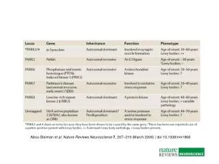
Download
1 / 15
150 likes | 251 Views
Cellular Neuroscience (207) Ian Parker. Lecture # 4 - The Hodgkin-Huxley Axon. http://parkerlab.bio.uci.edu. An electrical depolarization that propagates rapidly (up to 10s of m per sec) along nerve axons. The Action Potential. record. stimulate. + 50 mV. overshoot. 0 mV.

E N D
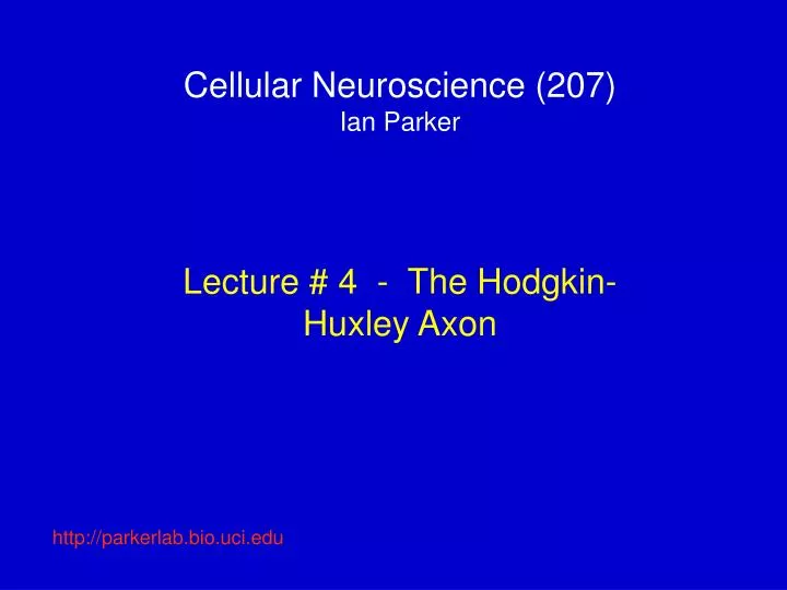
Cellular Neuroscience (207)Ian Parker Lecture # 4 - The Hodgkin-Huxley Axon http://parkerlab.bio.uci.edu
An electrical depolarization that propagates rapidly (up to 10s of m per sec) along nerve axons The Action Potential record stimulate + 50 mV overshoot 0 mV depolarization Rising phase Falling phase repolarization - 70 mV hyperpolarization Afterpotential (undershoot) Stimulus artifact Conduction delay
Basic mechanisms of the action potential The action potential is a brief time when the membrane potential is ‘flipped’ – positive rather than negative inside. This arises because the cell membrane becomes transiently permeable to Na+ ions, which rush into the cell down their concentration gradient, depolarizing it toward ENa. RISING PHASEFALLING PHASE • Na channels inactivate, so depolarizing, inward Na+ current stops. • Voltage-gated K channels open • Efflux of K+ ions down their electrochemical gradient repolarizes the cell toward Ek • Repolarization causes K channels to shut, but slow gating may cause ‘undershoot’ below normal resting potential. 1.Depolarization (e.g. excitatory synaptic input) opens voltage-dependent Na channels. 2. Na+ ions enter cell causing… 3. Depolarization
Some properties of the action potential • 1. Action potentials are all-or-none events. Once a stimulus exceeds threshold (ca. -45 mV) an action potential is triggered. Size of the action potential (peak ~ + 50 mV) is fixed, and does not depend on stimulus strength. • 2. Action potentials propagate without decrement at a finite speed. Speed is fast by biological standards (several m per sec vs. um per sec for ‘chemical;’ signals), but much (million-fold) slower than an electrical signal along a wire. • 3. Refractory period. After one action potential there is a short time (ms) when an axon cannot be stimulated to give another action potential. Primarily due to the time for Na channels to recover from inactivation. This is important because it; • a. Stops action potentials from traveling ‘backwards’ • b. Sets a limit to the maximum frequency of action potentials an axon can transmit.
Hodgkin - Huxley analysis of the action potential (early 1950s) Voltage-clamp Technique that allows the voltage across an axon membrane to be held at any desired level, while measuring the resulting current flow across the membrane. Used with giant (1 mm diameter) squid axon, that allows easy insertion of intracellular electrodes. Feedback circuit – compares the actual membrane potential with the desired command voltage. Any difference (error) is amplified and inverted, and fed back into the axon as a current to bring the potential to the desired level (like cruise control on a car). Current flowing from the circuit thus gives a direct measure of current flowing acros the axon membrane.
Currents flowing across a squid axon in response to voltage steps Depolarization to voltages more positive than about -25 mV evokes a complex series of currents. A transient current –usually inward-, followed by a slower developing , maintained outward current. The initial transient current at first becomes larger (more inward current) with increasing depolarization, then reduces to zero at ~ +60 mV, and inverts to become outward at yet more positive voltages. The slower current is always outward, and becomes increasingly large at more positive potentials. Depolarization to -35 mV evokes only passive responses Hyperpolarization evokes only passive, leakage currents depolarize or hyperpolarize Resting potential
How to make sense of this – pharmacologically dissect the transient and maintained currents into their ionic components Total currents evoked by a range of depolarizing stimuli Blocking Na channels with tetrodotoxin abolishes the initial transient current, leaving only the slower, maintained outward K current. Blocking K channels with TEA abolishes the slow outward current, leaving just the fast, inward Na currrent
Current/voltage relationships for the initial and maintained current components Current amplitudes measured at their peaks The delayed, outward current increases progressively at increasingly positive voltages Both currents begin to activate at about -35 mV. The initial transient current increases with voltages up to about +20 mV, then declines to zero at about +50 - +60 mV, and becomes outward at voltages > +60 mV
Currents through Na and K channels reflect both the Ohmic dependence of current flow through single channels, and the voltage-dependence of channel open probability We can separate these two effects by calculating whole-cell conductance as a function of voltage e.g. for transient Na current IM ENa = +50 mV Sigmoid relationship reflecting voltage-dependent activation of Na channels Current across the axon membrane is the product of the single-channel Na current and the number of Na channels open at a given voltage. We can estimate the latter by calculating Na conductance; gNa = IM/VM-ENa 0 mV VM g Na Ichannel VM VM 0 +50 mV -50 mV The conductance/voltage relationship for K channels looks very similar, except that the initial ‘turn-on’ is a little less steep
Equivalent circuit diagram for an axon membrane The Na and K channels can be thought of as variable resistors, whose values depend on voltage, and which determine the importance of their respective ‘batteries’ (Na and K equilibrium potentials) in setting the final voltage across the cell membrane. Changing the membrane potential involves charging the membrane capacitance, so the voltage changes during an action potential depend on the time course (kinetics) of the Na and K conductance changes as well as their peak values.
So, what are the kinetics of gNa and gK? 0 mV VM -60 mV gNa gK During depolarization, gNa shows both time-dependent activation and inactivation. gK shows only activation. During the falling phase of an action potential, gK declines because the membrane potential repolarizes, NOT because K channels inactivate The kinetics of Na channel activation and inactivation, and the kinetics of K channel activation all become faster at more positive potentials - 20 mV +20 mV gK gNa
VM Sigmoid rise Exponential decay gK Time course of K channel activation and closing H-H expained the openinng of a K channel as being controlled by movement of several independent ‘particles’ (voltage sensors). The channel is open only if all are in the ‘ON’ position. Suppose 4 particles, each with probability n of being in the ON position. Probability of channel opening is then given by n4 Further suppose that probability n changes exponentially with time following a voltage step VM gK (varies as n) n gK (varies as n4)
More about gating particles The K channel molecule has 4 charged ‘particles’ that move according to the voltage across the membrane. [In the 1950s these particles were merely postulates – we now know they correspond to the S4 regions of the channel molecule] - - - - - - - - - - - - - Out + + + + + + + + + + + + + + ON position OFF position + + + + + + + + + + + + + + In - - - - - - - - - - - - - n varies with voltage and with time. H-H characterized it by two parameters; ninfinity probability of being in the ON state after holding at a given voltage for a very long time tn the rate at which n changes following a step to a new voltage. From their experimental data H-H could derive empirical values for these parameters.
What about Na channels? H-H described Na channel activation in the same way as for K channels, by movement of gating particles. For the Na channel, these are referred to as m (not n), and movement of only 3 (not 4) was required to give the best fit to the data. Also, another gating particle (h – only one per channel) was introduced to account for the inactivation of the Na channel
The Hodgkin Huxley Equation Ionic currents across the axon membrane can be described in terms of three components; IM = m3hgNa(E-ENa) + n4gK(E-EK) +gL(E-EL) Na current K current ‘leak’ current [Don’t worry; you wont be asked to remember this in an exam! ] All of the electrical excitability of the membrane is embodied in the time- and voltage-dependence of n, m and h. The model accurately predicts observed action potentials in many species, and is one of the few cases where we can reduce biology to an equation. But, like any other model it cannot prove the existence of underlying mechanisms.