Download
1 / 34
340 likes | 640 Views
EVALUATION. Clinical – History & Physical Laboratory Hemodynamic All parameters are indirect, nonspecific measures of volume Serial evaluations necessary ≈ fluid therapy Modalities should complement one another. PHYSICAL. Most reliable preoperatively

E N D
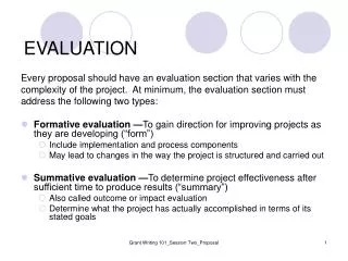
EVALUATION • Clinical – History & Physical • Laboratory • Hemodynamic • All parameters are indirect, nonspecific measures of volume • Serial evaluations necessary ≈ fluid therapy • Modalities should complement one another
PHYSICAL Most reliable preoperatively Skin turgor, hydration of mucous membranes, fullness of peripheral pulse, capillary refill, resting HR & BP and changes from the supine to sitting or standing position, urinary excretion and fontanels in babies.
LABORATORY Serial hematocrits Arterial blood pH Urinary specific gravity/osmolality >1.01/450 mOsm/kg Serum blood urea nitrogen (BUN)-to-creatinine ratio > 10:1 Indirect indices of volume, esp intraoperatively Only X-ray signs reliable measures of volume overload – Kerly B lines or intestitial markings
HEMODYNAMIC CENTRAL VENOUS PRESSURE (CVP) Cardiac output is based on the Frank starling mechanism where force of contraction is determined by the initial fiber length and the contractility of cardiac muscle to determine stroke volume We do not measure stroke volume, so pressure is used as a surrogate The placement of a central venous catheter with its tip at junction SVC & RA provides measurable parameter of volume status or preload of patient
PULMONARYARTERY PRESSURE In the normal individual CVP measurement provides a reasonably accurate estimate of the filling pressures of both R & L atria. In some situations not, and infusion of fluids or inotropic agents titrated against CVP may not result in optimum cardiac function LV failure with pulmonary oedema Interstitial pulmonary oedema of any cause Chronic pulmonary disease Valvular heart disease
PULSE PRESSURE VARIATION Ventilation causes changes in intrathorasic pressure, influences cardiac filling Responsible variation in BP during ventilation Identify highest and lowest BP Subtract highest DBP from highest SBP and lowest DBP from lowest SBP Render pulse pressure variation Divide diff btw HPP & LPP by mean X 100
Highest SBP = 100 mmHgHighest DBP = 60 mmHgLowest SBP = 90 mmHgLowest DBP = 55 mmHgHPP = 100 – 60 = 40 mmHgLPP = 90 - 55 = 35 mmHgDifference btw HPP & LPP = 40 – 35 = 5 mmHgMean PP = (40+35) / 2 = 37.5 mmHgtherefore the PPV = (5/37.5) x 100 = 13.3%> 12% indication of hypovolaemia ~ respond fluidvolume, < 8% non-responders, 8-12 grey area
INTRAVENOUS FLUIDS Crystalloids, Colloids or both • Crystalloids ~ aqueous sol low-molecular-weight ions (salts) ± glucose • Colloids ~ high-molecular-weight sub :- Protein colloids – Albumin :- Non protein colloids – gelatins (haemaccel, gelofusin) hydroxyethylstarchs (voluven, venofundin) sugars (dextrans)
Replacing intravascular volume deficit with crystalloids ~ 3X volume needed using colloids • Intravascular fluid deficits ~ more rapidly corrected using colloid solutions • Surgical patients ~ extracellular fluid deficit > intravascular deficit • Rapid administration of large amounts of crystalloids (>4-51) more frequently associated with significant tissue oedema • Intravascular ½ life crystalloids 20-30 min, colloids ½ life 3-6 hours
PERIOPERATIVEFLUID THERAPY • Replacement: pre-existing deficits, maintenance requirements and surgical wound losses • Maintenance fluid requirements • 70 kg person fasting for 8h amounts to: (40 + 20 + 50) ml/h X 8h = 880 ml
BLOOD • Volume • Oxygenation • Clotting
Prem 95ml / kg • Neonate 90ml / kg • > 3month 80ml / kg • > 1y 70ml / kg • MABL = EBV × ( I Hct – F Hct) / Mean Hct Mean Hct = ( I Hct + F Hct ) / 2 Whole blood = ( F Hct – I Hct) × Kg × 2.5 Packed RBC = (F Hct – I Hct) × Kg × 1,5
DO2 = CO × CaO2 = (70 × 72) × [ (Hb × 1,34 × SaO2 ) + (0,031 × PaO2) ] = 5 × 200ml = 1000ml/min • Extraction ~ 200ml/min
COMPLICATIONS OF BLOOD TRANSFUSIONS HEMOLYTIC REACTIONS Involves specific destruction of transfused RBC by recipient’s antibodies, less common – hemolysis recipient’s RBC due to transfusion of antibodies ACUTE HEMOLYTIC REACTIONS Fatal 1:100000 In awake patients – chills, fever, nausea, chest and flank pain. In anaesthetized pts -↑ temp, ↑HR, hypotension, hemoglobinuria and diffuse oozing in surgical field. Disseminated intravascular coagulation & renal shutdown Severe with as little as 10-15ml ABO-incompatibility
MANAGEMENT OF REACTION • Hemolytic reaction suspected ~ stop transfusion • Recheck identity bracelet against blood slip • Draw blood for Hb, compatibility, platelet count & coagulation studies • Urinary catheter inserted & urine checked for Hb • Osmotic diuresis initiated with mannitol & iv fluids • Presence of rapid blood loss – Platelets & FFP
DELAYED HEMOLYTIC REACTIONS • Following ABO & Rh-compatible transfusion, 1 – 1.6% chance antibodies against Kell, Duffy, Kidd etc antigens • Extravascular hemolysis • Mild – malaise, jaundice & fever 2-21 days after • Treatment primarily supportive
NONHEMOLYTIC REACTIONS Sensitization of the recipient to donor white cells, platelets or plasma proteins • Febrile reactions • Urticarial reactions • Anaphylactic reactions • Noncardiogenic pulmonary oedema • Graft-Versus-Host disease • Posttransfusion purpura • Immune suppression
ANAPHYLAXIS Definition: Allergic condition which results from an antibody-antigen reaction rapidly after the antigen entered the systemic circulation. Signs: Resp: Bronchospasm, laryngeal oedema CVS: Circulatory collapse – hypotension Skin: Wheel & flare
Management Initial Therapy - Stop drug - Call for help - Airway management - Feet elevation - Drug Rx Adrenaline: 0.5 – 1mg IMI/10min 50 – 100μg IVI/ 1min (hypotension)
Secondary Therapy - Antihistamines - Corticosteroids - Catecholamine infusion Adrenaline 0.05 – 0.1 μg/kg/min - Blood gas acidosis consider Bicarbonate 0.5-1 mmol/kg - Airway evaluation before extubation - Bronchodilators in persistent bronchospasm
Investigations: -Only after emergency treatment has been completed - Dx on clinical grounds - Bloods: Serum tryptase concentration 1 hour after reaction 10ml red top centrifuge and store @ -20ºC until send to Lab - Patient and GP must be alerted toward the reaction and drug causing it. Potentially
OBESITY BMI– Body Mass Index is the weight (kg) divided by the square of the height (m) Normal range 18-25 Overweight >27 Obese >30 Morbid obesity >35 Massive morbid obesity >40 ? modeling not <18 Broca Index - normal weight (kg) = height (cm) minus 100 for males or 105 for females - children weight (kg) = 10 + 2 age
Distribution – truncal, buttocks • Respiratory - Difficult intubation • - FRC • - work of breathing, chest compliance • - risk of aspiration: gastric volume, : Hiatus hernia :intra- abdominal pressure - Obstructive sleep apnea esp after GA or opioids, PCA ~ safer • Nocturnal CPAP nasal oxygen mask • Apnea monitor
Cardiovascular - blood volume and CO • Difficult: IV access : BP measurement (cuff size –20% > arm diameter ) arterial line • PCA better than IM opiods • Tromboprophylaxis & mobilization post-op • Medical conditions – Diabetes mellitus, Cushing’s syndrome, hypothyroidism, syndromes (Prader-Willi or Lawrence-Moon-Biedl) • Table max 150kg
LARYNGOSPASM Definition – Acute glottic closure by the vocal cords Presentation – Crowing or absent inspiratory sounds and marked tracheal tug Differential diagnosis - Bronchospasm - Laryngeal trauma / airway oedema - Recurrent laryngeal nerve damage - Tracheomalacia - Inhaled foreign body - Epiglottitis or croup
Management • Avoid painful stimuli • Remove irritants from airway • 100% oxygen • CPAP mask, jaw thrust • ? Deepen anaesthesia • Intractable: Muscle relaxation and intubation
PACEMAKERS Indications– Third degree heart block Mobitz type II block Trifassicular block: RBBB : Left ant/post hemiblock : First degree heart block Sick sinus syndrome Symptomatic bradycardia Post MI, HOCM, torsade de pointes
Pacemaker code • Position 1- chamber(s) paced • Position 2- chamber(s) sensed • Position 3-response to a sensed elect. Signal • Position 4- rate modulation • Position 5- multi-site pacing
Anaesthetic implications - Follow up clinic, function - Pre-op ECG : Absence of all spikes may represent appropriate sensing or total failure! - Loss of capture : Hypokalaemia After defibrillation MI over lead Toxic levels of local anaesthetic Lead dislodgement - Bipolar diathermy safe - MRI