Download
1 / 2
20 likes | 34 Views
A lymphangiogram is a special x-ray of the lymph nodes and lymph vessels.<br><br>The lymph nodes and vessels are not seen on a normal x-ray, so a dye is injected into the body to highlight the area being studied.<br><br>The skin over your groin will be numbed and then a thin needle is inserted under ultrasound guidance into a lymph node in your groin. Contrast will be injected through the needle and into the lymph node.

E N D
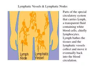
Lymphangiography And LymphaticInterventions A lymphangiogram is a special x-ray of the lymph nodes and lymphvessels. The lymph nodes and vessels are not seen on a normal x-ray, so a dye is injected into the body to highlight the area beingstudied. The skin over your groin will be numbed and then a thin needle is inserted under ultrasound guidance into a lymph node in your groin. Contrast will be injected through the needle and into the lymph node. A type of x-ray machine, called a fluoroscope, projects the images on a TV monitor. The provider uses the images to follow the dye as it spreads through the lymphatic system up your legs, groin, and along the back of the abdominal cavity to identify the site of lymphatic leak. Then, embolizing agent (blocking agent) is used to block the site ofleak. Why(Indications)? Provides structural and functionalinformation regarding the lymphaticsystem. Primarily indicated in patients with chylousleaks. Also helps in identifying site of lymphatic obstruction, abnormal lympho-lymphatic, or lymphovenousconnections. Why Not(Contraindication)? These include pulmonary insufficiency and a right to left cardiacshunt. What you are to do beforeprocedure(Preparation)? • Visit us in OPD for assessment of varicocele with ultrasound. Get lab investigation (*PT/INR, Serum Creatinine, Viral markers) done and book yourappointment • Get admission in day care on scheduled time and date with 4-6 Hoursfasting • If you are on blood thinner like Aspirin inform duringappointment. • One accompanyingperson • Need to sign a consent form forprocedure
Approx. Stay inhospital? We have very fast and competent working team (Consultant, fellow, clinical assistant, technician and ward assistant) which provide you comfortable atmosphere and ease your nerves. Usual time of stay is around 2-3days. Complications: • Mild pain/burningsensation • Infection • Failure oftherapy • Resume towork? • You can resume your work after 5-7 days, if the existing diseaseallows. • Results? • Success rate is approx. 80-90% • Lymphedema • Diagnosis • If you're at risk of lymphedema — for instance, if you've recently had cancer surgery involving your lymph nodes — your doctor may diagnose lymphedema based on your signs andsymptoms. • If the cause of your lymphedema isn't as obvious, we may order imaging tests to get a look at your lymph system. Tests mayinclude: • MRI scan. Using a magnetic field and radio waves, an MRI produces 3-D, high-resolution images. • CT scan. This X-ray technique produces detailed, cross-sectional images of your body's structures. CT scans can reveal blockages in the lymphatic system. • Doppler ultrasound. This variation of the conventional ultrasound looks at blood flow and pressure by bouncing high-frequency sound waves (ultrasound) off red blood cells. Ultrasound can help findobstructions. • Radionuclide imaging of your lymphatic system (lymphoscintigraphy). During this test you're injected with a radioactive dye and then scanned by a machine. The resulting images show the dye moving through your lymph vessels, highlightingblockages.