Download
1 / 4
40 likes | 50 Views
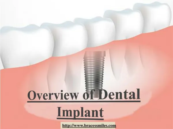
The successful rehabilitation of edentulous and partially dentate situation with dental implant has been well documented in the recent years. Despite a high success rate, implants failure is inevitable. Dental implant related complication can cause significant problems due to the difficulty in removing the fractured implant and the resultant ridge defects, in addition needing to modify the prosthetic appliance. This paper provides an overview on various techniques available to remove failed dental implant.

E N D
Journal Home Page www.bbbulletin.org BRITISH BIOMEDICAL BULLETIN Review Article An Insight into Dental Implant Removal: An Overview Manu Rathee*1, Mohaneesh Bhoria1 and Shashikala Jain2 1Department of Prosthodontics, Post Graduate Institute of Dental Sciences, Pt. B.D Sharma University of Health Sciences, Rohtak, Haryana, India 2Department of Prosthodontics, Surendera Dental College and Research Institute, Sriganganagar, Rajasthan, India A R T I C L E I N F O Received 12 Feb. 2015 Received in revised form 22 Mar. 2015 Accepted 24 Mar. 2015 Keywords: Dental Implants, Implant Failures, Endoscope, Implant Complication. Corresponding author:Department of Prosthodontics, Post Graduate Institute of Dental Sciences, Pt. B.D Sharma University of Health Sciences, Rohtak, Haryana, India. E-mail address: ratheemanu@gmail.com A B S T R A C T The successful rehabilitation of edentulous and partially dentate situation with dental implant has been well documented in the recent years. Despite a high success rate, implants failure is inevitable. Dental implant related complication can cause significant problems due to the difficulty in removing the fractured implant and the resultant ridge defects, in addition needing to modify the prosthetic appliance. This paper provides an overview on various techniques available to remove failed dental implant. ©2015 British Biomedical Bulletin. All rights reserved
Ratheeet al_____________________________________________________ ISSN-2347-5447 Introduction Implant therapy reported a high success rate in the literature.1 Among various complications implant fracture has been reported as a rare complication. A literature review described biological and technical complications during a follow-up period of 5 years after implant therapy. Implant fracture was reported as a complication that may occur with an incidence of <1%during 5-year follow-up.2 Mostly, when a fracture occurs, the implant left ‘submerged’ inside the alveolar bony crest and another suitable anatomic location identified and implants were placed. However, under individual clinical scenario implant may require removal, such as the fracture of the unretrievable internal screw for abutment retention, an untreatable peri- implantitis, the malpositioning of an implant in the arch, and when treatment does not provide patients with satisfactory function and aesthetics. The removal of implants that is well osseointegrated may be performed in different ways.3 This article provides an overview of available technique of implant removal. Implant failure and complications This involves etiology.4,5 Causes related to early failure includes overheating of the bone during osteotomy preparation, over preparation of the osteotomy, implant during surgery, poor bone quality, lack of primary stability, and macro-motion caused by overload or parafunction. Implants with any of the above mentioned etiologies are usually mobile, and early removal is easily performed. Late failure is most often related to peri-implantitis, occlusal trauma, implant fracture or implant overload. Many late- failing implants remain partially integrated with no mobility. Implants that fail due to peri-implantitis show many clinical signs similar to those found around periodontally diseased teeth. These signs include bleeding on probing, suppuration, pain, increased probing depth, radiographic evidence of bone loss, presence of periopathogens bacteria, and site-specific However, unlike teeth with moderate or advanced periodontitis, many implants with peri-implantitis do not display mobility. Factors necessary for successful integration of dental implants have been proposed. Of these, a lack of mobility is a primary prerequisite. However, even when implants are non-mobile, indications for their removal may exist, such as fracture, malposition, infection, pain, and implantitis. Although several reviews of the literature cite various techniques to treat implants with peri-implantitis removal, these unpredictable. In cases of advanced peri- implantitis or implant fracture, removal of the affected implant is usually necessary.6 Techniques of implant removal Many of these non-mobile implants require surgical intervention for removal. Methods of implant removal include: 1.Counter-torque (CTRT) It is the least invasive technique for removing an implant without damaging surrounding structures. Cases amenable to removal with a counter-torque ratchet. The use of a counter-torque ratchet should be considered the option of choice if the implant is able to be engaged and reverse- torqued until mobile.3,4 2.Bone removal techniques (BRTs) Piezo tips, elevators, forceps, and trephine burs. These may involve: (a)Apicoectomy The apicoectomy approach can be used to retrieve implant fragments not infections. advanced peri- without appear techniques ratchet technique multifactorial contamination high-speed burs, BBB[3][1][2015] 124-127
Ratheeet al_____________________________________________________ ISSN-2347-5447 visible in the alveolar arch and to preserve bone for the placement of new implants. The apicoectomy technique is a surgical root approach used to preserve the tooth. Here, the prime consideration knowledge as correct localization of the root apex is required. Radiographic investigation help to localize the fractured implant and a lateral bone window is made to permit good visualization of the bone condition while avoiding excessive destruction of the bone, which is usually a consequence of other techniques. (b)Implant removal from maxillary sinus The anatomical approximation of maxillary sinus often poses problems for the implant placement in the posterior maxillary region. This is especially evident with sinus pneumatisation that reaches just a few millimeters above the crest of the alveolar ridge. Placement of immediate implants under such clinical scenario may create unexpected problems communication with the maxillary sinus and poor bone quality that might lead to the displacement of the implant into the sinus. Many approaches suggested one approach was to leave it, as long as no complications arose. However, due to development of a severe infection around the implant in the sinus, two main treatment modalities have been proposed for the removal of foreign bodies in the sinuses (1) an intraoral approach with the creation of a bony window in the anterior-lateral wall of the maxillary sinus, which can be used endoscopically as well; and (2) a transnasal approach with functional endoscopic sinus surgery. Management of this complication can addressed with the aid of endoscopy.8,9 (c)Trephine bur technique The use of a trephine is the most common procedure for the removal of fractured implants or for histological evaluations. utilization of a circular drill with diameter larger than the implant diameter to remove the osseointegrated implant. However, if replacement with a new implant is required in the same location, the external diameter of the drill must be taken into account, to insert an implant with a larger diameter to ensure primary stability. Disadvantages include the removal of copious quantities of bone to overcome osseointegration, leading to the destruction of surrounding bone. The other involves is the difficulty of removal when the implants are not visible clinically. All these techniques, however, will result in the removal of part of the surrounding bone, possible overheating of the bone and spreading of titanium particles into the adjacent tissues. Furthermore, there may be damage to adjacent vital structures, such as roots, Schneiderian membrane and vascular- nervous bundles.10-13 (d)Edentulous ridge expansion (ERE) This technique can be utilized to remove failed implants due to peri- implantitis, fracture or malpositioning. Advantages include a minimally invasive procedure, reduction of the loss of surrounding bone, overheating of the bone and reduction in the spreading of titanium particles into the adjacent tissues. This technique allowed a safer approach to structures, such as roots, Schneiderian membrane and vascular-nervous bundles when compared to the use of trephines. Here, blade was used to separate the buccal bony wall from the implant surface, providing a ‘bone flap’ and subsequently, a lever was applied to remove the implant. However, when this technique is applied care must be provided to avoid damage to the surrounding bone and the fracture of the buccal bony plate.14 This technique involves is anatomic the force of of undetected reduction of the adjacent anatomic BBB[3][1][2015] 124-127
Ratheeet al_____________________________________________________ ISSN-2347-5447 6. Sanchez-Garces Perimplantitis. Med Oral Patol Oral or Bucal. 2004; 9(suppl):69-74; 63-69. Caccioli P. Apicectomy: localization and isolation of the radicular apex. Acta Biomed Ateneo Parmense. 1992; 63:97-100. Nakamura N, Mitsuyasu T, Ohishi M. Endoscopic removal of a dental implant displaced into the maxillary sinus:Technical note. Int J Oral Maxillofac Surg 2004; 33:195–7. Iezzi G, Scarano A, Mangano C, Cirotti B, Piattelli A. Histologic results from a human implant retrieved due to fracture 5 years after insertion in a sinus augmented with anorganic bovine bone. J Periodontol 2008; 79:192–8. 10.Degidi M, Iezzi G, Scarano A, Piattelli A. Immediately loaded titanium implant with a tissue-stabilizing/maintaining (beyond platform switch) retrieved from man after 4 weeks: histomorphometrical evaluation. A case report. Clin Oral Implants Res 2008; 19:276–82. 11.Degidi M, Perrotti V, Strocchi R, Piattelli A, Iezzi G. Is insertion torque correlated to bone-implant contact percentage in the early healing period? histomorphometrical evaluation of 17 human retrieved dental implants. Clin Oral Implants Res 2009; 20:778–81. 12.Iezzi G, Degidi M, Scarano A, Perrotti V, Piattelli A. Bone response to submerged, unloaded implants inserted in poor bone sites: a histological and histomorphometrical study of 8 titanium implants retrieved from man. J Oral Implantol 2005; 31:225–33. 13.Scarano A, Degidi M, Iezzi G, Petrone G, Piattelli A. Correlation between implant stability quotient and bone-implant contact: a retrospective histomorphometrical study of seven titanium implants retrieved from humans. Clin Implant Dent Relat Res 2006; 8:218–22. 14.Vercellotti T. Piezoelectric surgery in implantology: a piezoelectric ridge expansion technique. Int J Periodontics Restorative Dent 2000; 20:358– 65. MA, Gay-Escoda C. Conclusion Regular review and maintenance are essential to maintain the health of implant supporting tissues and to prevent related complications. However, it is important to realise that complications do occur and for patients to appreciate the value of long-term care. Once dental implants have been removed, they are manufacturer for replacement, depending on the company’s protocol. It is essential that radiographic and photographic evidence is taken before the dental implants are returned as well as for medico-legal reasons. Source (s) of support Nil. Conflict of interest Nil. References 1. Branemark P-l, Zarb GA, Albrektsson T. Tissue Integrated Osseointegration in Clinical Dentistry. Chicago, III: Quintessence; 1985. 2. Eckert SE, Merav SJ, Cal E, Ovj RK. Analysis of incidence and associated factors with fractured implants: a retrospective study. Int J Oral Maxillofac Implants. 2000; l5:662-567. 3. Froum S, Yamanaka T, Cho SC, Kelly R, James SS, Elian N, Techniques to Remove a Failed Integrated Implant. Compendium of Continuing Education in Dentistry. 2011; 32(7):22-30. 4. Gealh WC, Mazzo V, Barbi F, et al. Osseointegrated implant fracture: Causes and treatment. J Oral Implantol. 2011; 37:499– 503. 5. Rangert B, Krogh PH, Langer B, Van Roekel N. Bending overload and implant fracture: a retrospective clinical analysis. Int J Oral Maxillofac Implants. 1995; 10: 326- 334. 7. 8. returned to the 9. design a histological and Prostheses: A histological and histological and case report-a new BBB[3][1][2015] 124-127