Download
1 / 76
830 likes | 1.19k Views
Spinal Tumors Good, Bad, and Ugly. Spinal Metastases. Sohail Bajammal, MB ChB September 18, 2006 Hamilton General Hospital – Weekly Spine Rounds. Permission to use for educational uses with reference to the source. 1 st Objective. To recognize red flags and avoid delayed diagnosis.

E N D
Spinal TumorsGood, Bad, and Ugly Spinal Metastases Sohail Bajammal, MB ChB September 18, 2006 Hamilton General Hospital – Weekly Spine Rounds Permission to use for educational uses with reference to the source
1st Objective To recognize red flags and avoid delayed diagnosis
2nd Objective Most of what you need to know for the Royal College Exam
Things to cover • Characteristics • Presentation • Evaluation • Treatment
Tumors of the Spine • Vertebral tumors: • Primary: 2-5% • Primary benign • Primary malignant • Benign more common than malignant • Secondary (metastases): 95-98% • Spinal cord tumors (neurosurgery): • Extra-dural • Intra-dural: • extra-medullary • intra-medullary
Tips for Differential • Young patients (20s-30s) more like to be benign tumors (except osteosarcoma and Ewing’s sarcoma) • Benign lesions tend to favor posterior elements
Osteoid Osteoma • Age: 1st 3 decades, peak 15 • 10-25% of osteoid osteoma occur in spine • 70% of painful juvenile scoliosis are due to osteoid osteoma • Pain, worse at night, responds to NSAIDs
Osteoid Osteoma • Investigations: • X-ray: nidus surrounded by a halo, scoliosis • Bone scan: most sensitive, target sign • CT: most specific • Treatment: • Medical: NSAIDs, observation • Surgical: if progressing or not responding • Excision • Percutaneous radiofrequency ablation
Osteoblastoma • 40% of osteoblastoma involves spine • Histologically similar to osteoid osteoma, but larger in size, different presentation • Clinical Presentation: • Pain, activity, not as responsive to NSAIDs • Larger size (>2cm) cortical expansion radiculopathy • Scoliosis: less common than osteoid osteoma
Osteoblastoma • Investigations: • X-rays: more readily detected • Treatment: • Slowly progressive less likely non-operative • Surgical: • Curettage: 15% recurrence, 50% in high grade • Marginal resection: less recurrence • Malignant transformation: very rare
Giant Cell Tumor (GCT) • Characteristics: • 5-10% of GCT involves spine • Most common in sacrum • Vertebral body • Clinical Presentation: • Age: 30s-40s, more in women • Variable: slowly growing to locally aggressive with metastases • Delayed of diagnosis • Pain and radiculopathy
Giant Cell Tumor (GCT) • Investigations: • X-rays: well-demarcated, radiolucent lesion with cortical expansion and local remodeling • Treatment: • En bloc resection: optimal, but higher morbidity • Curettage: acceptable option, higher recurrence • Prognosis: • Poorer than GCT in appendicular skeleton • Recurrence: 80% in stage III • Metastases to lung: 10%
Osteochondroma • <10% of all osteochondroma • More in cervical • If multiple hereditary multiple exostoses • Slowly growing rare mechanical or compressive symptoms • Treatment: • Mainly observation • Resection: if symptomatic
Eosinophilic Granuloma (EG)Langerhans Cell Histiocytosis • Benign, self-limiting process of well-demarcated bone resorption, ? etiology • 1st – 2nd decade, Male 2:1 • Spine involved in 10-15% of EG • Common sites: skull, pelvis, ribs, shoulder • Associated with 2 systemic diseases: • Hand-Schüller-Christian disease • Letterer-Siwe disease
Eosinophilic Granuloma (EG)Langerhans Cell Histiocytosis • Investigations: • Spine X-rays: Vertebra plana (D/D) • Skeletal survey • Abdominal U/S: hepatosplenomegaly (HSC) • Treatment: • Observation: because self-limiting • Surgical resection: if progressive kyphosis or progressive neurological symptoms • Low dose radiotherapy: if not amenable for surgery
Vertebra Plana (FETISH) • Fracture • Eosinophilic granuloma • Tumor • Metastases • Myeloma • Ewing’s • Osteosarcoma • ABC • Infection • TB • Osteomyelitis (disc involvement) • Steroids • Hemangioma
Aneurysmal Bone Cyst (ABC) • Characteristics: • Spine involved in 10-30% of ABC • Posterior element of thoracolumbar spine • May involve multilevel adjacent segments • 1st – 2nd decade • Investigations: • X-rays: cortical expansion and thinning, “bubbly” appearance • MRI: fluid/fluid level
Aneurysmal Bone Cyst (ABC) • Treatment Options: • Curettage • Wide local excision • Embolization • Radiation • Prognosis: • Recurrence: 15-30%
Hemangioma • Characteristics: • Most common tumor of the spine • Commonly incidental finding • 10% of autopsy • Single lesion in 2/3 of cases • Mainly in vertebral body, thoracic spine • Clinical Presentation: • Neural compression by cortical or soft tissue expansion
Hemangioma • Investigations: • X-rays: • Able to detect only if involves 30-40% of body • Vertical trabecular striations like a honeycomb • CT or MRI: for subtle lesions • Treatment Options: • Low dose radiation • Embolization • Surgical resection & stabilization: if instability • Vertebroplasty and kyphoplasty
Differential Diagnosis(Anterior Spine) • Non tumor • Infection (discitis) • TB • Benign • Neurofibroma • Hemangioma • GCT • EG • ABC (more posterior) • Malignant • Metastases • Myeloma
Differential Diagnosis(Posterior Spine) • Benign • Osteochondroma • Osteoblastoma • Osteoid osteoma • ABC • Malignant • Metastases
Osteosarcoma • 3-14% of malignant tumors of spine • 2% of all osteosarcoma in the body • Mainly in vertebral body, lumbosacral • Bimodal age: • 10 – 25 yr: primary • Older than 50yr: secondary (radiation, Paget’s) • Many histological types • Poorer prognosis and older age than appendicular osteosarcoma
Osteosarcoma • Treatment: • Neoadjuvant chemotherapy surgical resection Adjuvant chemotherapy • If not amenable for surgical resection: chemo and radiotherapy • Bad prognostic factors: • Metastases at diagnosis • Large size • Sacral location • Intralesional resection
Chondrosarcoma • Characteristics: • 2nd most common primary malignant bone tumor (after chordoma) • 7-12% of all spine tumors • Age: 40s, more in men • Treatment: • Resistant to radiotherapy and chemotherapy • Surgical excision
Chordoma • Characteristics: • Most common primary malignant tumor of spine (excluding lymphoproliferative disorders) • Age: 50s – 60s, Males: 3x more common • Remnants of the primitive notochord midline • Sacrococcygeal > Base of skull > V. body (C) • Clinical Presentation: • Gradual onset, disregarded, Pain, numbness, weakness, constipation or incont. • Sacrococcygeal lesions palpable by DRE
Chordoma - Diagnosis • X-rays: midline, lytic or mixed lytic and blastic • CT: check involvement of local structures (rectum, vessels) • MRI: check involvement of dura & roots • Biopsy: posterior midline, never trans-rectal • Histology: lobular framework of physaliphorous cells
Chordoma - Treatment • Highly resistant to chemo and radiotherapy • Radiotherapy for positive margins or palliative • Lesions above S3: usually requires anterior and posterior approach for excision • Unilateral retention of all roots: near normal bowel, bladder, and sexual function • Sacrificing S2 incontinent • Metastases: liver, lungs, lymph nodes, peritoneum
Multiple Myeloma • Characteristics: • B-cell lymphoproliferative diseases • Rapidly progressive and highly lethal (20% survival at 5 yr) • Age: 60s – 70s • Investigations: • X-ray: looks normal; Bone scan: cold • CT and MRI: delineate lesion • Serum and urine protein electrophoresis • 20% of cases: only urine is positive
Multiple Myeloma - Treatment • Very radiosensitive main modality • Chemotherapy for systemic component • Bracing: for lesion <50% of vertebral body • Surgery indicated for: • Stabilization of the spine • Decompression of neurological elements • Local control if recurrence or no response to radiation therapy • Follow with MRI and serum electrophoresis
Significance • The spine is the most common site for skeletal metastases • Metastatic lesions are the most common tumors of the spine (95-98%) • Vertebral body affected first • Approximately 70% of patients who die of cancer have evidence of vertebral metastases on autopsy Harrington 1986
Common Primary Sites • Breast (21%) • Lung (14%) • Prostate (7.5%) • Renal (5%) • GI (5%) • Thyroid (2.5%)
Level of Metastases • Thoracolumbar 70% • Lumbosacral 20% • Cervical 10%
Clinical Presentation • Pain (85%) Hyperemia, expansion, nerve compression, cord compression, pathologic fractures & instability • Weakness (34%) Spinal cord compression in 20% • Mass (13%) • Constitutional Symptoms
History • Age: high level of suspicion • Details of the pain: insidious or acute, ± trauma, axial, ± radiculopathy, unrelenting, non-mechanical, worse at night, change in features if chronic • Personal history of cancer • Constitutional symptoms • Review of systems: thyroid, breast, chest, GI, GU & skin • Any age-specific screening tests by GP • Social history: smoking, alcohol, exposure to carcinogen • Family history of malignancy
Physical Exam • Thorough examination of thyroid, breast, lung, abdomen, pelvis, prostate, skin, lymph nodes (referrals) • Spine: • Look: alignment • Feel: focal tenderness • Move: ROM • Neurological examination: gait, power, sensation, reflexes (DTR, abdominal, Babinski, Hoffman), clonus
Evaluation • History • Physical Exam • Laboratory: • CBC, ESR, CRP, Lytes, BUN, Creatinine • Ca, PO4, Alk Phosph • Urinalysis: routine, Bence-Jones Proteins • Special: PSA, thyroid Fxn, serum and urine protein electrophoresis, liver function tests, stool guaiac, CEA • Radiological • Biopsy
Radiological Evaluation • Local: • X-ray of spine: AP, lateral, oblique • “winking owl” sign: pedicle destruction • Vertebral body destruction is not visible until 30-50% of trabeculae are involved • Negative x-ray does not rule out tumor • Bone Scan: screening, cold in MM • CT: bony architecture • MRI + gadolinium: gold standard
Radiological Evaluation • Staging: • CT chest, abdomen and pelvis with oral and IV contrast • Bone Scan • Mammogram
Biopsy • Indicated if primary diagnosis is unclear after workup: • Remote history of malignancy with long disease-free interval • Options: • CT-guided: most accessible lesion, minimal morbidity, tattoo tract for later excision • Open: cost, delay, definitive for benign tumors • Culture every tumor and biopsy every infection
Goal of Management Maximize quality of life
To achieve the goal…. • Provide pain relief • Improve or maintain neurologic function • Restore or maintain the structural integrity of the spinal column
Options of Treatment • Orthotic • Steroids • Radiotherapy • Chemotherapy • Hormonal Therapy • Surgery • Combination Multi-disciplinary approach
Pitfall Aggressive chemotherapeutic regimens for patients with spinal pain not responding to conventional therapy without ruling out subtle mechanical etiology Severe depression of bone marrow that surgery or radiotherapy are no longer feasible
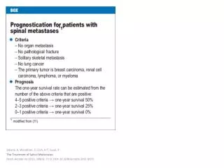
Decision Making (Prognostic Decision Rules) • Frankel et. al. Paraplegia 1969 • Harrington JBJS(A) 1986 • Tokuhashi et. al. Spine 1990 • Tomita et. al. Spine 2001