Download
1 / 29
310 likes | 421 Views
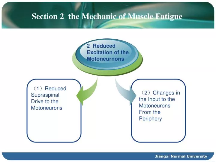
( 1 ) Reduced Supraspinal Drive to the Motoneurons. ( 2 ) Changes in the Input to the Motoneurons From the Periphery . Section 2 the Mechanic of Muscle Fatigue. 2 Reduced Excitation of the Motoneurnons. 2 Reduced Excitation of the Motoneurnons.

E N D
(1)Reduced Supraspinal Drive to the Motoneurons (2)Changes in the Input to the Motoneurons From the Periphery Section 2 the Mechanic of Muscle Fatigue 2 Reduced Excitation of the Motoneurnons Jiangxi Normal University
2 Reduced Excitation of the Motoneurnons • Several factors contribute to reduced excitation of spinal motorneurous. Basically ,any reduction in the excitatory drive and/or increase in inhibitory influence acting on the motoneurons, whether from subpraspinal levels, from the segmental circuitry, or from the periphery, will reduce firing rates. Jiangxi Normal University
(1)Reduced Supraspinal Drive to the Motoneurons • Not only acute reduction in activity but also long-term reductions, such as during limb immobilization, have been reported to disproportionately reduce maximal voluntary force compared with twitch and tetanic forces. This may be taken to indicate that voluntary muscle activity is necessary to retain the ability to produce high levels of drive to the motoneuron pool . In a way ,the reduced drive following a period of inactivity can be regarded as the inverse of neural adaptation to strength training . A similar explanation may be offered for the greater prevalence of central fatigue in older subjects reported by Bilodeau et al.(2001). • Change in the human motor cortex have been shown to accompany central fatigue. Experiments with transcranial magnetic stimulation(TMS)in healthy humans have shown decreased intracortical facilitation after fatiguing exercise, and the changes were contined to the areas of the motor cortex involved in the fatiguing contractions. The mechanisms behind these changes could not be established, although events upstream of the motor cortex have been considered to be possible candidates(Tergau et al.2000), such as supraspinal effects of group Ⅲand Ⅳ muscle afferents (Gandevia 1998). As alluded to previously, this means that elements of central fatigue could originate in the periphery. Jiangxi Normal University
Although little is known about the neurophysiological or biochemical mechanisms behind central fatigue ,some theories exist, one of which is the so-called branched-chain amino acid (BCAA) theory. The main points of this theory are as follows: 1、BCAAS and tryptophan enter the brain via the same amino carrier, and competition between the two types of amino acid for entry into the brain can occur. 2、In the brain ,tryptophan is converted to the neurotransmitter 5-hydroxytryptamin(5-HT), also known as serotonin, which is known to promote sleep and tiredness, a fact that makes it a possible fatigue-promoting substance. 3、During physical activity ,BCAAs are taken up by muscle, shifting the balance between the plasma concentrations of tryptophan and BCAAs in the direction of trytophan. This can increase the uptake of tryptophan in the brain and the subsequent formation of 5-HT(Newsholme and Blomstrand 1995). So far ,however ,this remains a theory because experiments involving ingestion of tryptophan and BCAAs during sustained exercise have failed to show a performance effect (Van Hall et al.1995). Jiangxi Normal University
(2)Changes in the Input to the Motoneurons From the Periphery • In conscious humans ,fatiguing muscle contractions are accompanied by a decreased in the firing rates of a motoneurons. This has been interpreted as a mechanism to ensure proper matching between motoneuron firing rate and muscle unit force output and is commonly referred to as muscle wisdom • During a fatiguing maximal voluntary contraction ,the electromyographic activity declines roughly in parallel with the loss of force. This decline has been attributed to reflex inhibition of the motoneuron pool . • Oligosynaptic spinal pathways are likely contributors , possibly mediated by group Ⅲ(corresponding to group A) orⅣ(corresponding to group C) muscle afferents, which are known to be activated by muscle contractions . Group Ⅳ contains unmyelihated nerve fibers responsive to pain stimuli, whereas group Ⅲ are thin, myelinated fibers responsive to pain and temperature stimuli. Neuropharmacological research has shown that some group Ⅲ and Ⅳ muscle afferents—the so called capsaicin-sensitive nerves-are responsive to protons as well. There is a good correlation between tissue PH and afferent discharge when the PH falls to 6.6 or less , and the resulting inhibition of motoneurons can be regarded as a protective mechanism. Jiangxi Normal University
(2)Changes in the Input to the Motoneurons From the Periphery • however , there was not a parallel decline in motorneuronal firing rate , the activation of motor units would become supratetanic, and rate coding as a means of force modulation would be ineffective . this is the reason why the phenomenon has been named muscle wisdom. • Muscle spindles are the main source of afferent excitation from the periphery , and spindle afferent activity declines during muscle contraction . Blockade of spindle afferents reduces motoneuron firing rates , indicating that fusimotor-mediated spindle facilitation is necessary to reach peak rates . Although spindle afferent activity declines during static contractions, resulting in a disfacilitation of a motoneurons (figure 15.6), the situation could be different during dynamic contractions. During concentric work , fusimotor drive is insufficient to compensate for muscle shortening, and spindle afferent become silent. In eccentric contractions ,on the other hand , the stretch sensitivity of muscle spindles increase, which increase the ability in spindle afferents and increase muscle stiffness . Jiangxi Normal University
(1)Motor Axon propagation Failure- Branch Failure (2)Fatigue at the Neuromuscular Junction 3 Factors Affecting Generation of an Endplate Potential Jiangxi Normal University
(1)Motor Axon propagation Failure- Branch Failure • It has been known for quite some time that action potentials can fail to propagate along each branch of a motor axon , and that failure is likely to occur at axonal branching points. Experiments have suggested that shifts in the ionic balance over the axon membrane could be responsible (figure 15.7). such ionic perturbations are more likely to occur in smaller axons , because the surface-to-volume ration is larger in thin axons than in thicker ones, and they are more likely to occur during high-frequency stimulation. Consequently ,branch failure is believed to start peripherally and spread gradually in a centripetal direction. Jiangxi Normal University
(1)Motor Axon propagation Failure- Branch Failure Jiangxi Normal University
(1)Motor Axon propagation Failure- Branch Failure • The most peripheral branch points are the terminal branchings of the axon as it approaches each • Muscle fiber (cf. figure 3.9). For each individual axonal branch ,the effect of the ionic perturbations on action potential propagation is probably transient, meaning that an affected synaptic terminal within the motor endplate is not completely silenced, but that occasional action potentials are missing, decreasing the firing rate for that particular terminal at the level of muscle fiber and reducing the safety factor of the motor endplate in question. • A branch failure where the axon divides into separate branches for each muscle fiber will inevitably reduce impulse frequency to the muscle fibers innervated by the affected branches and will correspondingly reduce the force contribution of those muscle fibers Jiangxi Normal University
(2)Fatigue at the Neuromuscular Junction • The successful transmission of an action potential in a motor endplate depends on the size of the combined EPSPs (excitatory postsynaptic potentials, see chapter 4) of all the synaptic terminals of the synaptic terminals of the endplate in relation to the threshold for muscle fiber action potential generation. • Theoretically , a transmission failure at the neuro-muscle junction may be the result of each of the following factors or a combination of them: a reduced number of discharging synaptic terminals , a reduced number of synaptic vesicles in the discharging terminals, a reduced sensitivity of the Ach receptors (AChRs) in the postsynaptic membrane (figure 15.8). Jiangxi Normal University
(2)Fatigue at the Neuromuscular Junction Jiangxi Normal University
(2)Fatigue at the Neuromuscular Junction • A reduced number of discharging synaptic terminals in a motor endplate can result from a branch failure within the terminal arborization of the axon , as outlined previously. To a certain extent, this can be handled by the safety factory. The safety factor for type Ⅱ muscle fibers has been reported to be larger than for type Ⅰ fibers, but for both fiber types it is large enough to ensure reliable neuromuscular transmission under nonfatigued conditions. On repetitive stimulation, however, the safety factor for type fibers remains unchanged, whereas that of type Ⅱ fibers rapidly declines . The reason for this difference is so far speculative. It is also unknown how and to what extent the ionic perturbations created by repeated muscle fiber action potentials influence propagation in the neighboring terminal branches of the axon. • During postnatal growth , both muscle fibers and motor endplates increase in size , the latter relatively more than the former, thus increasing the size of motor endplates in relation to muscle fiber size. In accordance with this ,newborn rats have been found to be more susceptible to neuromuscular transmission failure than adult animals. In line with this , the relative increasing in the size of endplates seen in animals subjected to training can be taken as an indication that training confers resistance to neuromuscular transmission failure. For natural reasons , corresponding findings in humans are lacking . Jiangxi Normal University
Summary • Muscle fatigue can be categorized as central or peripheral, but it should be acknowledged that the words central and peripheral are not used in the anatomically correct way. The main reason for this are that the definition of central fatigue is operational, and that factors leading to central fatigue may have their origin in the periphery. Central fatigue is said to be present when maximum voluntary force is less than maximal evocable force. The interpolated twitch technique is the main method to separate central from peripheral fatigue. • Transcranial magnetic stimulation has revealed the presence of decreased intracortical facilitation after fatiguing exercise, and the changes are confined to the somatotopically relevant parts of the motor cortex. The changes, however ,could be secondary to changes in afferent activity to the motor cortex. Jiangxi Normal University
Summary • For normal ,rested muscle , the launching of an action potential from the motoneuron is the point of no return in muscle control. Thus, in nonfatigued muscle, any change in the balance between excitatory and inhibitory inputs to the motoneurons , in favor of latter, will reduce the impulse frequency to the muscle units and tend to lower their force input. This can be attributed to reduced supraspinal drive or reduced muscle spindal afferent actibity. Theoretically ,failure of propagation of the nerve action potential (brance failure) and neuromuscular transmission failure also can be give rise to central fatigue, but the practical importance of this is probably limited, at least in daily life. Jiangxi Normal University
4 Fatigue Attributable to Processes Beyond the Neuromuscular Junction-Peripheral Fatigue (1)Failure in the propagation of the muscle fiber action potential-high-frequency fatigue (2)Fatigue at the T-Tubule- Sarcoplasmic Reticulum Junction-Low-Frequency Fatigue (3)Myofibrillar Fatigue Jiangxi Normal University
(1)Failure in the propagation of the muscle fiber action potential-high-frequency fatigue • Indirect evidence indicates that failure in the propagation of muscle action potentials along the T-tubules plays an important role, and this has been attributed to accumulation of K+ in the T-tubule lumen. Theoretically, the T-tubules would be expected to be the more susceptible to ionic perturbations because of repetitive stimulation. First , the lumen is very small in relation to the T-tubule membrane. Second ,the density of Na+-K+ ATPase molecules(the sodium-potassium pump) has been claimed to be lower in the T-tubule membrane than in the surface membrane, and the more important effects of an increased extracellular K+ concentration have been considered likely to involve excitation-contraction coupling via the T-tubules , rather than the excitability of the surface membrane. However , human studies provided support for the possible role of T-tubules in high-frequency fatigue(HFF), because a kind of fatigue resenmbling HFF result from stimulation of human tibialis anterior muscle under ischemic conditions. Muscles held at shorted length during stimulation fatigued more than muscles held at optimum length. Both groups recovered substantially after cessation of stimulation, although still ischemic, but the muscle fatigue in the shortened position showed further recovery when returned to optimum length. These results are consistent with restricted movement and consequent accumulation of K+ in the lumen of T-tubules, possibly because of narrowing of their openings to the surface of the fibers when in the shortened position. Jiangxi Normal University
(1)Failure in the propagation of the muscle fiber action potential-high-frequency fatigue • Indirect support for a propagation failure in the T-tubules comes from the demonstration in freeze-clamped fatigued muscle fibers that centrally located myofibrils had a wavy appearance, indicating that they were less activated and thus longer than more peripherally located myofibrils. • HFF is induced by high-frequency stimulation, and it has been questioned whether HFF is a physiologically relevant and “normal” type of fatigue, mainly because the frequencies necessary to induce HFF are higher than those thought to occur during normal sustained isometric contractions. On the theoretical basis, it has been calculated that Na+-K+ ATPase is able to keep pace with the influx of sodium ions and efflux of potassium at excitation frequencies up to around 55Hz . Jiangxi Normal University
(2)Fatigue at the T-Tubule- Sarcoplasmic Reticulum Junction-Low-Frequency Fatigue • In 1997, Richard Edwards and coworkers described a type of muscle fatigue that they called low-frequency fatigue (LFF).it was characterized by being most pronounced at low frequencies (figure 15.9) and by having a very slow recovery –in fact taking hours, maybe days ,despite absence of any signs of metabolic or electrical disturbance in the muscle. the slow recovery is in sharp contrast to the very fast recovery is in the sharp contrast to the very fast recovery , in the order of minutes, seen after HFF. Jiangxi Normal University
(2)Fatigue at the T-Tubule- Sarcoplasmic Reticulum Junction-Low-Frequency Fatigue Jiangxi Normal University
(2)Fatigue at the T-Tubule- Sarcoplasmic Reticulum Junction-Low-Frequency Fatigue • The slow recovery indicated that some kind of repair process involving protein synthesis was necessary , and this suspicion was strengthened by the fact that LFF is common after eccentric contractions , a type pf muscular activity known to cause substantial damage. • LFF was accompanied by a uniform reduction of cytosolic calcium across the fatigue muscle fiber, whereas no change was seen in calcium sensitivity or in maximal calcium-activated tension, thus settling the previously mentioned uncertainty. It remained unknown, however, what was responsible for the reduced calcium levels. Furthermore, although decreased cytosolic calcium has been shown during LFF, it may not be the only cause ; a redistribution of sarcomere lengths also has been suspected to contribute to LFF. Binder-Macleod and Russ presented evidence in support of two factors contributing to LFF:one rapidly recovering , metabolite-dependent mechanism, and a slow-developing, slow-recovering factor, which is not the result of any metabolite buildup. Jiangxi Normal University
(3)Myofibrillar Fatigue • Fatigue caused y inability of the muscle fiber to react properly to elevated cytosolic Ca2+ concentration is called myofibrillar fatigue. In such cases , the force/Ca2+ concentration curve is shifted to the right ,but this has been shown only for type Ⅰfiber. Under these circumstances , caffeine-induced elevation of cytosolic Ca+2 concentration is unable to increase force (figure 15.11). • In normal , rested muscle , the force per sarcomere is proportional to the number of active across-bridges. Theoretically ,a reduced force output despite unchanged sarcomere length and cytosolic Ca2+ concentration can be attributable to a reduced number of active cross-bridges or a reduced force per cross-bridge. Jiangxi Normal University
(3)Myofibrillar Fatigue Jiangxi Normal University
(3)Myofibrillar Fatigue • Fatigability of Different Fiber Types:There are typical different in the fatigability of human muscle fiber types. TypeⅠfibers are the more fatigue resistant, type ⅡX the least, and type ⅡA intermediate. This is demonstrated very clearly by figure 15.12, which shows the correlation between percentage of type ⅡX fibers in human quadriceps muscle and peak torque decline after 50 maximal knee extensions . The open circles in figure 15.12 denote individuals with less than 35% type Ⅱ fibers, that is, with more than 65% typeⅠ. Part of the variation also could be attributable to the fact that differences between “identical” fiber types in different individuals are even larger than difference between different fiber types within one muscle. this is caused by differences in individuals’ training status and can even apply to difference between muscles in different parts of the body in one individual, depending on his or her training level. Jiangxi Normal University
(3)Myofibrillar Fatigue Jiangxi Normal University
(3)Myofibrillar Fatigue • Basically, the different fatigue resistance of muscle fiber types derives from three characteristics: contractile economy, oxidative capacity, and relative training status, which among other things is subsequent to their rank in the recruitment hierarchy. • The relative oxidative potential of the different muscle fiber types is typeⅠ>typeⅡ>typeⅡX, which also contributes to the differences in fatigue resistance between the muscle fiber types. Because the oxidative potential of a muscle fiber is highly trainable, the fatigue resistance of low- threshold typeⅠmuscle fibers may be attributable in part to their recruitment history. The size principle governs the recruitment of motor units during isometrix and concentric work. In these cases, the recruitment hierarchy can be likened to a flight of stairs, which has to be climbed from the bottom every time, regardless of how high you have to climb. Jiangxi Normal University
Summary • Peripheral fatigue is the kind of fatigue that is not alleviated by direct stimulation of the muscle by the twitch interpolation technique. It can be caused by factors affecting the propagation of muscle action potentials along the surface membrane and into the T-tubules, factors affecting the release of calcium from the sarcoplasmic reticulum, or factors affecting the response of individual myofibrils to increase levels of cytosolic calcium. • Propagation failure is mostly seen after unphysiologically high stimulation frequencies and seems to be of limited importance in daily life. Such HFF is rapidly reversed when the fatiguing activity is terminated, and it is believed to be caused by ionic perturbations, in particular a loss of K+ from the cytosol to the extracellular space. Ionic perturbations in the T-tubule are often claimed to be responsible for failure of T-tubule are often claimed to be responsible for failure of T-tubule function, but direct evidence is lacking. Jiangxi Normal University
Summary • Failure in the calcium release process gives rise to a characteristic force deficit, which is more pronounced at low than at high stimulation frequencies, hence the name LFF. In contrast to HFF,LFF shows a very slow recovery, indicating that molecules critically involved in the calcium release process are seriously damaged and in need of replacement . the mechanism behind this process is not known yet, but elevated levels of calcium are implicated, phenomenon referred to as the calcium paradox. • Inability of the individual myofibrils to respond properly to increased levels of cytosolic calcium is called myofibrillar fatigue. It can be caused by a decreased number of active across-bridges or a decreased force per active across-bridge. • TypeⅠis the most fatigue-resistant muscle fiber type, typeⅡX is most easily fatigue. This differences is based on three characteristics: muscle fiber contractile economy, oxidative capacity, and relative training status, subsequent to their rank in the recruitment hierarchy. Jiangxi Normal University