Download
1 / 72
720 likes | 1.18k Views
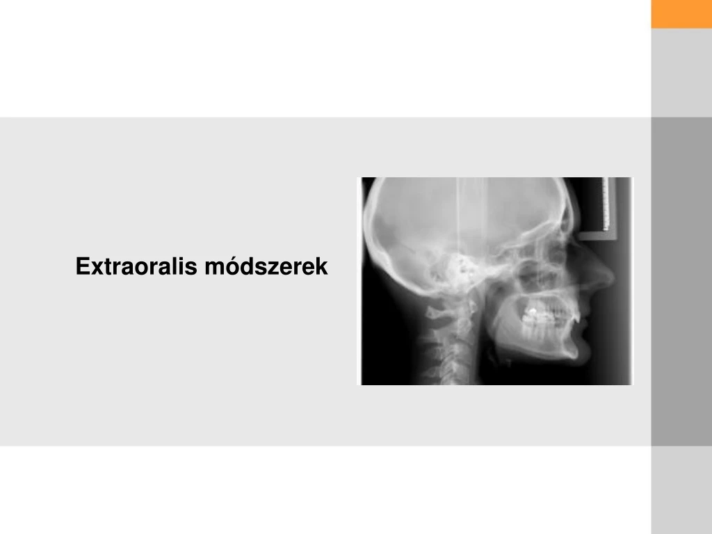
Extraoralis módszerek. BEVEZETÉS -. A film és a röntgencső is extraorális elhelyezkedésű. Nagyobb terület vizsgálata. Pontatlanabb,mint az intraorális felvétel. Koponya és ízület vizsgálata. Indikáció: Trauma Impaktált fog Állkapocs ízületi probléma Fejlődési rendellenesség.

E N D
BEVEZETÉS- • A film és a röntgencső is extraorális elhelyezkedésű. • Nagyobb terület vizsgálata. • Pontatlanabb,mint az intraorális felvétel. • Koponya és ízület vizsgálata. • Indikáció: • Trauma • Impaktált fog • Állkapocs ízületi probléma • Fejlődési rendellenesség
Erősítő fólia • Fluoreszkáló anyagot tartalmaz. • Előnyök: kevesebb sugárterhelés kevesebb mozgási műtermék • Alkalmazás -extraoralis film -digitális felvétel
Fontosabb extraoralis projekciók • Standard occipitomental 0º • 30º Occipitomental • Posteroanterior (Koponya) • Lateralis és kefalometriai felvétel (Koponya) • Posteroanterior (Állcsont) • Reverse Towne’s • Submentovertex
As Low As Reasonably Achievable
Indikáció: • Középarc törés • Proc. Coronoideus törés • Sinus Maxillaris, Frontalis és Ethmoidal sejtek Standard occipitomental 0º
Indikáció: • Középarc törés • Proc. Coronoideus törés • Sinus Maxillaris, Frontalis 30º Occipitomental
Posteroanterior (Koponya) Indikáció: • Koponyatörés • Homlokboltozat vizsgálata • Egyéb megbetegedések: Morbus Paget Myeloma multiplex Hyperparathyroidizmus • Intracranialis kalcifikáció
Lateralis és kefalometriai projekció (Koponya) • Koponyalap törés • Felső állcsont törés • Sinus frontalis , maxillaris és sphenoidalis vizsgálata • Egyéb megbetegedések: Morbus Paget Myeloma multiplex Hyperparathyroidizmus • Sella Turcica: Hypophysistumor
Kefalometria • Koponyaméréstan. • Kefamoletriai analízisek által normától való eltérés meghatározása. • Dentális vagy szkeletális eltérések megkülönböztetése.
PA Water’s view (PNS) Indikation: • Sinus maxillaris • Sinus frontalis
Reverse Towne’s Indikáció: • Condylus törése • TMJ
Submentovertex Indikációk: • Koponyaalap és palatum lézió • Sinus sphenoidalis • Arcus zygomaticus törés
Rétegvastagság -Front 9 mm -Molare16 mm
Panoráma- Indikáció • Összkép megítélése • Tályog • Cysta • Számbeli és alakbeli rendellenesség • Tumor • Idegentest • Retinált fog • Mesiodens • TMJ
Panoráma-TMI Indikáció- • Ízületi megbetegedés • Proc. Condyleus mutáció - Aszimmetria • Erozió • Nagy Osteophyta • Tumor vagy törés.
Egyéb módszerek • CBCT • CT • MRI • UH
Cone-Beam CT Introduced to the US in 2000 • 2002 (~10) • 2003 (~30) • 2004 (~75) • 2005 (~175) • 2006 (~350) Rapid adoption in dentistry • Dental Schools • Dentists, Specialists, Imaging Centers • Cone-beam computed tomography (CBCT) is a recent technology initially developed for angiography in 1982 and subsequently applied to maxilofacial imaging.
Cégek: • AFP Newtom • Hitachi : Mercuray • Image Science International / Danaher: ICAT • Imtec / Kodak : Iluma • Morita: Accuitomo • Planmeca: ProMax 3D • Sirona: Galileos • Vatech : DCT& VCT • Yoshida /Terarecon: FineCube
Cone Beam Maxillofacial Imaging Systems Newtom - 3G Scanner Vatech - VCT Planmeca – Promax 3D Sirona - Galileos
Alkalmazás: Arc-Állcsont –Szájsebészet Fogászat Fül-Orr-Gégészet • PIXEL→VOXEL
C B C T versus Medical C T • Med CT • Conventional linear fan beam • Single row or a series (4, 8, 12, 32, 64) of solid state detectors • Provides a set of consecutive slices of the patient • CBCT • Cone beam • Square 2 dimensional array of detectors • Provides a volume of data
CONVENTIONAL CT X-ray source Fan’ of X-rays Detector
CBCT/CBVT X-ray source Cone’ of X-rays Detector Detector
CBCT Ebene: Axial Sagittal Transaxial Coronal
Axial (Transverse) This is an Axial image.. …that represents this area of anatomy
Coronal Coronal Plane slices through the anatomy from side to side. Click
Sagittal Sagittal Plane is a slice through the anatomy from front to back Click
Series of Cross-Sectionals/Transaxials Cross sectional images of an area can be developed with .5 to 5mm spacing between images.
Clinical Applications of CBCT • Dental Implant Planning & Guidance • Temporomandibular Evaluation • Pre-surgical Assessment • Impacted Teeth • Reconstructive • Airway Assessment • Orthodontic Assessment • Periodontics • Endodontics • Pathology
Clinical Applications of CBCT • Dental Implant Planning & Guidance • Temporomandibular Evaluation • Presurgical Assessment • Impacted Teeth • Reconstructive • Airway Assessment • Orthodontic Assessment • Periodontics • Endodontics • Pathology
-Dental Implants Clinical Applications of CBCT