Download
1 / 1
20 likes | 170 Views
Frequency of MDM2 Amplification in Malignant Peripheral Nerve Sheath Tumors: Non-Correlation with Tumor Grade, Cellularity and MIB1 Proliferation Index Michelle L. Wallander 1 , Sheryl Tripp 1 and Lester J. Layfield 2

E N D
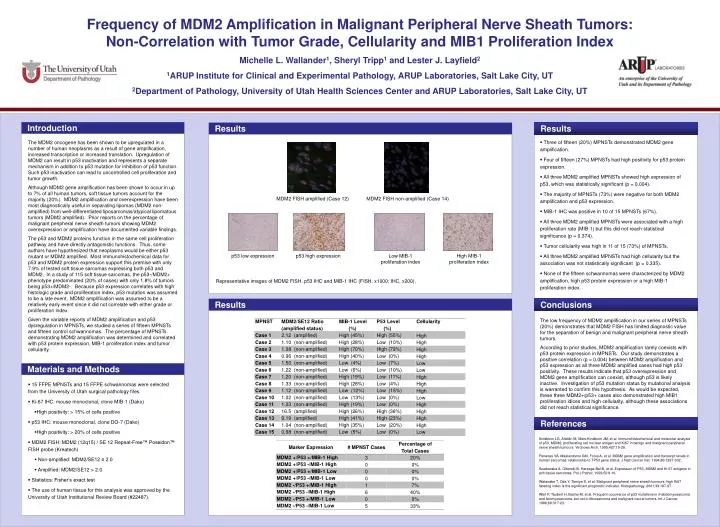
Frequency of MDM2 Amplification in Malignant Peripheral Nerve Sheath Tumors: Non-Correlation with Tumor Grade, Cellularity and MIB1 Proliferation Index Michelle L. Wallander1, Sheryl Tripp1and Lester J. Layfield2 1ARUP Institute for Clinical and Experimental Pathology, ARUP Laboratories, Salt Lake City, UT 2Department of Pathology, University of Utah Health Sciences Center and ARUP Laboratories, Salt Lake City, UT Introduction Results Results • Three of fifteen (20%) MPNSTs demonstrated MDM2 gene amplification. • Four of fifteen (27%) MPNSTs had high positivity for p53 protein expression. • All three MDM2 amplified MPNSTs showed high expression of p53, which was statistically significant (p = 0.004). • The majority of MPNSTs (73%) were negative for both MDM2 amplification and p53 expression. • MIB-1 IHC was positive in 10 of 15 MPNSTs (67%). • All three MDM2 amplified MPNSTs were associated with a high proliferation rate (MIB-1) but this did not reach statistical significance (p = 0.374). • Tumor cellularity was high in 11 of 15 (73%) of MPNSTs. • All three MDM2 amplified MPNSTs had high cellularity but the association was not statistically significant (p = 0.335). • None of the fifteen schwannomas were characterized by MDM2 amplification, high p53 protein expression or a high MIB-1 proliferation index. The MDM2 oncogene has been shown to be upregulated in a number of human neoplasms as a result of gene amplification, increased transcription or increased translation. Upregulation of MDM2 can result in p53 inactivation and represents a separate mechanism in addition to p53 mutation for inhibition of p53 function. Such p53 inactivation can lead to uncontrolled cell proliferation and tumor growth. Although MDM2 gene amplification has been shown to occur in up to 7% of all human tumors, soft tissue tumors account for the majority (20%). MDM2 amplification and overexpression have been most diagnostically useful in separating lipomas (MDM2 non-amplified) from well-differentiated liposarcomas/atypical lipomatous tumors (MDM2 amplified). Prior reports on the percentage of malignant peripheral nerve sheath tumors showing MDM2 overexpression or amplification have documented variable findings. The p53 and MDM2 proteins function in the same cell proliferation pathway and have directly antagonistic functions. Thus, some authors have hypothesized that neoplasms would be either p53 mutant or MDM2 amplified. Most immunohistochemical data for p53 and MDM2 protein expression support this premise with only 7.9% of tested soft tissue sarcomas expressing both p53 and MDM2. In a study of 115 soft tissue sarcomas, the p53-/MDM2+ phenotype predominated (20% of cases) with only 1.8% of tumors being p53+/MDM2-. Because p53 expression correlates with high histologic grade and proliferation index, p53 mutation was assumed to be a late event. MDM2 amplification was assumed to be a relatively early event since it did not correlate with either grade or proliferation index. Given the variable reports of MDM2 amplification and p53 dysregulation in MPNSTs, we studied a series of fifteen MPNSTs and fifteen control schwannomas. The percentage of MPNSTs demonstrating MDM2 amplification was determined and correlated with p53 protein expression, MIB-1 proliferation index and tumor cellularity. MDM2 FISH amplified (Case 12) MDM2 FISH non-amplified (Case 14) p53 low expression p53 high expression Low MIB-1 proliferation index High MIB-1 proliferation index Representative images of MDM2 FISH, p53 IHC and MIB-1 IHC (FISH, x1000; IHC, x200). Conclusions Results The low frequency of MDM2 amplification in our series of MPNSTs (20%) demonstrates that MDM2 FISH has limited diagnostic value for the separation of benign and malignant peripheral nerve sheath tumors. According to prior studies, MDM2 amplification rarely coexists with p53 protein expression in MPNSTs. Our study demonstrates a positive correlation (p = 0.004) between MDM2 amplification and p53 expression as all three MDM2 amplified cases had high p53 positivity. These results indicate that p53 overexpression and MDM2 gene amplification can coexist, although p53 is likely inactive. Investigation of p53 mutation status by mutational analysis is warranted to confirm this hypothesis. As would be expected, these three MDM2+/p53+ cases also demonstrated high MIB1 proliferation idices and high cellularity, although these associations did not reach statistical significance. Materials and Methods • 15 FFPE MPNSTs and 15 FFPE schwannomas were selected from the University of Utah surgical pathology files. • Ki-67 IHC: mouse monoclonal, clone MIB-1 (Dako) • High positivity: > 15% of cells positive • p53 IHC: mouse monoclonal, clone DO-7 (Dako) • High positivity: > 20% of cells positive • MDM2 FISH: MDM2 (12q15) / SE 12 Repeat-Free™ Poseidon™ FISH probe (Kreatech) • Non-amplified: MDM2/SE12 ≤ 2.0 • Amplified: MDM2/SE12 > 2.0 • Statistics: Fisher’s exact test • The use of human tissue for this analysis was approved by the University of Utah Institutional Review Board (#22487). References Kindblom LG, Ahldén M, Meis-Kindblom JM, et al. Immunohistochemical and molecular analysis of p53, MDM2, proliferating cell nuclear antigen and Ki67 in benign and malignant peripheral nerve sheath tumours. Virchows Arch. 1995;427:19-26. Flørenes VA, Maelandsmo GM, Forus A, et al. MDM2 gene amplification and transcript levels in human sarcomas: relationship to TP53 gene status. J Natl Cancer Inst. 1994;86:1297-302. Szadowska A, Olborski B, Harezga-Bal B, et al. Expression of P53, MDM2 and Ki-67 antigens in soft tissue sarcomas. Pol J Pathol. 1999;50:9-16. Watanabe T, Oda Y, Tamiya S, et al. Malignant peripheral nerve sheath tumours: high Ki67 labeling index is the significant prognostic indicator. Histopathology. 2001;39:187-97. Würl P, Taubert H, Bache M, et al. Frequent occurrence of p53 mutations in rhabdomyosarcoma and leiomyosarcoma, but not in fibrosarcoma and malignant neural tumors. Int J Cancer. 1996;69:317-23. .