Download
1 / 34
360 likes | 778 Views
Lymphadenopathy. Soheir Adam , MD, MSc, MRCPath. The Lymphatic System.

E N D
Lymphadenopathy Soheir Adam , MD, MSc, MRCPath
The Lymphatic System • The body has approximately 600 lymph nodes, but only those in the submandibular, axillary or inguinal regions may normally be palpable in healthy people.1 Lymphadenopathy refers to nodes that are abnormal in either size, consistency or number. There are various classifications of lymphadenopathy, but a simple and clinically useful system is to classify lymphadenopathy as "generalized" if lymph nodes are enlarged in two or more noncontiguous areas or "localized" if only one area is involved.
Distinguishing between localized and generalized lymphadenopathy is important in formulating a differential diagnosis. • In primary care patients with unexplained lymphadenopathy, approximately 3/4 of patients will present with localized lymphadenopathy and 1/4 with generalized lymphadenopathy.
Lympahdenopathy • Findings from a Dutch study revealed a 0.6% annual incidence of unexplained lymphadenopathy in the general population. • Of 2,556 patients in the study who presented with unexplained lymphadenopathy to their family physicians, 256 (10 %) were referred to a subspecialist and 82 (3.2 %) required a biopsy, but only 29 (1.1 %) had a malignancy.
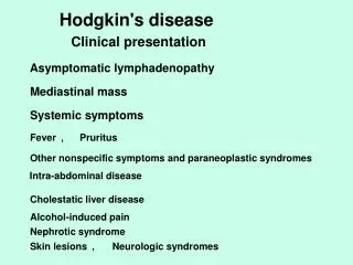
History • First, are there localizing symptoms or signs to suggest infection or neoplasm in a specific site? • Second, are there constitutional symptoms such as fever, weight loss, fatigue or night sweats to suggest disorders such as tuberculosis, lymphoma, collagen vascular diseases, unrecognized infection or malignancy?
History • Third, are there epidemiologic clues such as occupational exposures, recent travel or high-risk behaviors that suggest specific disorders? • Fourth, is the patient taking a medication that may cause lymphadenopathy? Some medications are known to specifically cause lymphadenopathy (e.g., phenytoin ), while others, such as cephalosporins, penicillins or sulfonamides, are more likely to cause a serum sickness-like syndrome with fever, arthralgias and rash in addition to lymphadenopathy.
Physical Examination • Size. • Pain/Tenderness:The presence or absence of tenderness does not reliably differentiate benign from malignant nodes. • Consistency: Stony-hard nodes are typically a sign of cancer, usually metastatic. Very firm, rubbery nodes suggest lymphoma. Softer nodes are the result of infections or inflammatory conditions. Suppurant nodes may be fluctuant. The term "shotty" refers to small nodes that feel like buckshot under the skin, as found in the cervical nodes of children with viral illnesses.
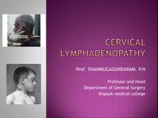
Physical Examination • Matting : can be either benign (e.g., tuberculosis, sarcoidosis) or malignant (e.g., metastatic carcinoma or lymphomas • Location: infectious mononucleosis causes cervical adenopathy and a number of sexually transmitted diseases are associated with inguinal adenopathy
Physical Examination • Supraclavicular lymphadenopathy has the highest risk of malignancy, estimated as 90 percent in patients older than 40 years and 25 percent in those younger than age. • Lymphadenopathy of the right supraclavicular node is associated with cancer in the mediastinum, lungs or esophagus. • The left supraclavicular (Virchow's) node receives lymphatic flow from the thorax and abdomen, and may signal pathology in the testes, ovaries, kidneys, pancreas, prostate, stomach or gallbladder. Although rarely present
Disorder Associated findings Test
Unexplained Lymphadenopathy Generalized Lymphadenopathy • almost always indicates a systemic disease is present, proceed with specific testing as indicated. • If a diagnosis cannot be made, the clinician should obtain a biopsy of the node. • The diagnostic yield of the biopsy can be maximized by obtaining an excisional biopsy of the largest and most abnormal node • The physician should not select inguinal and axillary nodes for biopsy, since they frequently show only reactive hyperplasia
Unexplained Lymphadenopathy Localized Lymphadenopathy • The decision about when to biopsy is more difficult. • Patients with a benign clinical history, an unremarkable physical examination and no constitutional symptoms should be reexamined in three to four weeks to see if the lymph nodes have regressed or disappeared. • Patients with unexplained localized lymphadenopathy who have constitutional symptoms or signs, risk factors for malignancy or lymphadenopathy that persists for three to four weeks should undergo a biopsy.
Unexplained Lymphadenopathy Localized Lymphadenopathy • Biopsy should be avoided in patients with probable viral illness because lymph node pathology in these patients may sometimes simulate lymphoma and lead to a false-positive diagnosis of malignancy.
Risk factors for NHL • immunosuppression or immunodeficiency • connective tissue disease • family history of lymphoma • infectious agents • ionizing radiation
Category Survival of untreated patients Curability To treat or not to treat Non-Hodgkin lymphoma Indolent Years Generally not curable Generally defer Rx if asymptomatic Aggressive Months Curable in some Treat Very aggressive Weeks Curable in some Treat Hodgkin lymphoma All types Variable – months to years Curable in most Treat A practical way to think of lymphoma
Diagnosis requires an adequate biopsy • Diagnosis should be biopsy-proven before treatment is initiated • Need enough tissue to assess cells and architecture • open bx vs core needle bx vs FNA
Stage I Stage II Stage III Stage IV Staging of lymphoma A: absence of B symptoms B: fever, night sweats, weight loss
Case: M.S. • 25 year old woman • persistent dry cough • fever, NS, weight loss x 3 months • left cervical lymphadenopathy (2 cm) • left supraclavicular node (2 cm) • no splenomegaly
Case: M.S. differential diagnosis • lymphoma • Hodgkin • non-Hodgkin • lung cancer • other neoplasms: thyroid, germ cell • non-neoplastic causes less likely • sarcoid, TB, ...
What next? • Needle aspirate of LN: a few necrotic cells • Needle biopsy of LN: admixture of B- and T-lymphocytes. A few atypical cells.
Case: M.S. staging investigations • CT chest / abdo / pelvis • bone marrow • gallium scan • Blood work: normal
Staging Investigations • bone marrow normal • CT scan: L supraclavicular adenopathy; large mediastinal mass; R hilum; no disease below diaphragm • gallium avid
What is her diagnosis and stage? • nodular sclerosis HD • stage IIB • with bulky mediastinal mass