Download
1 / 23
230 likes | 333 Views
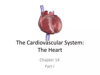
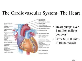
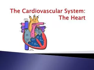
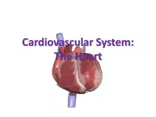
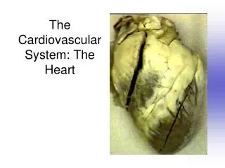
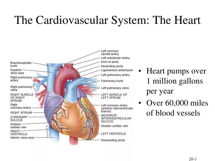
The Cardiovascular System: The Heart. Heart pumps over 1 million gallons per year Over 60,000 miles of blood vessels. Heart Location. Heart is located in the mediastinum area from the sternum to the vertebral column and between the lungs.

E N D
The Cardiovascular System: The Heart • Heart pumps over 1 million gallons per year • Over 60,000 miles of blood vessels
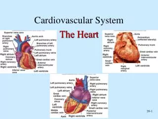
Heart Location • Heart is located in the mediastinum • area from the sternum to the vertebral column and between the lungs • Apex - directed anteriorly, inferiorly and to the left • Base - directed posteriorly, superiorly and to the right • Anterior surface - deep to the sternum and ribs • Inferior surface - rests on the diaphragm • Right border - faces right lung • Left border (pulmonary border) - faces left lung
Surface Projection of the Heart • Superior right point at the superior border of the 3rd right costal cartilage • Superior left point at the inferior border of the 2nd left costal cartilage 3cm to the left of midline • Inferior left point at the 5th intercostal space, 9 cm from the midline • Inferior right point at superior border of the 6th right costal cartilage, 3 cm from the midline
Pericardium • Fibrous pericardium • dense irregular CT • protects and anchors the heart, prevents overstretching • Serous pericardium • thin delicate membrane • contains • parietal layer-outer layer • pericardial cavity with pericardial fluid • visceral layer (epicardium) • Epicardium • visceral layer of serous pericardium • Myocardium • cardiac muscle layer is the bulk of the heart • Endocardium • chamber lining & valves
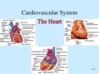
Surface anatomy • Sulci - grooves on surface of heart containing coronary blood vessels and fat • coronary sulcus • encircles heart and marks the boundary between the atria and the ventricles • anterior interventricular sulcus • marks the boundary between the ventricles anteriorly
Surface Anatomy • - posterior interventricular sulcus • marks the boundary between the ventricles posteriorly Posterior View
Chambers and Sulci • Four chambers • 2 upper atria • 2 lower ventricles
Semilunar valves open with ventricular contraction • allow blood to flow into pulmonary trunk and aorta • SL valves close with ventricular relaxation • prevents blood from returning to ventricles, blood fills valve cusps, tightly closing the SL valves Valves • A-V valves open and allow blood to flow from atria into ventricles when ventricular pressure is lower than atrial pressure • occurs when ventricles are relaxed, chordae tendineae are slack and papillary muscles are relaxed • A-V valves close preventing backflow of blood into atria • occurs when ventricles contract, pushing valve cusps closed, chordae tendinae are pulled taut and papillary muscles contract to pull cords and prevent cusps from everting
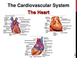
Right Ventricle Right Atrium • Receives blood from 3 sources • superior vena cava, inferior vena cava and coronary sinus • Interatrial septum partitions the atria • Fossa ovalis is a remnant of the fetal foramen ovale • Tricuspid valve • Blood flows through into right ventricle • has three cusps composed of dense CT covered by endocardium • Forms most of anterior surface of heart • Papillary muscles are cone shaped trabeculae carneae (raised bundles of cardiac muscle) • Chordae tendineae: cords between valve cusps and papillary muscles • Interventricular septum: partitions ventricles • Pulmonary semilunar valve: blood flows into pulmonary trunk
Left Ventricle Left Atrium • Forms the apex of heart • Chordae tendineae anchor bicuspid valve to papillary muscles (also has trabeculae carneae like right ventricle) • Aortic semilunar valve: • blood passes through valve into the ascending aorta • just above valve are the openings to the coronary arteries • Forms most of the base of the heart • Receives blood from lungs - 4 pulmonary veins (2 right + 2 left) • Bicuspid valve: blood passes through into left ventricle • has two cusps • to remember names of this valve, try the pneumonic LAMB • Left Atrioventricular, Mitral, or Bicuspid valve
Semilunar Valve(pulmonary) Interatrial septum Right atrium Tricuspid AV valve Semilunar Valve(aortic) Chordae tendinae Papillary muscle
Blood Circulation • Systemic circulation • left side of heart pumps blood through body • left ventricle pumps oxygenated blood into aorta • aorta branches into many arteries that travel to organs • arteries branch into many arterioles in tissue • arterioles branch into thin-walled capillaries for exchange of gases and nutrients • deoxygenated blood begins its return in venules • venules merge into veins and return to right atrium • Pulmonary circulation • right side of heart pumps deoxygenated blood to lungs • right ventricle pumps blood to pulmonary trunk • pulmonary trunk branches into pulmonary arteries • pulmonary arteries carry blood to lungs for exchange of gases • oxygenated blood returns to heart in pulmonary veins
Blood Circulation • Blood flow • blue = deoxygenated • red = oxygenated
SVC/IVCRight Atrium(tricuspid valve)Right Ventricle Passage of Blood through the Heart Body (pulmonary semilunar valve) Pulmonary Arteries Lungs Pulmonary Veins Left Atrium bicuspid (mitral) valve Body Left Ventricle Aorta (aortic semilunar valve)
The Heartbeat each heartbeat = cardiac cycle -SL valves close “dup” -AV valves open -filling of atria & ventricles begins -contraction of atria -AV valves open -filling of ventricles = “Ventricular Filling stage” -contraction of ventricles -AV valves close “lub” -SL valves open -blood to lungs and body **Heart Murmur**
Conduction System of Heart • Autorhythmic Cells • Cells fire spontaneously, act as pacemaker and form conduction system for the heart • SA node = 1. • cluster of cells in wall of Rt. Atria • begins heart activity that spreads to both atria • excitation spreads to AV node • AV node = 2. • in atrial septum, transmits signal to bundle of His • AV bundle of His = 3. • Divides into Right and Left Bundle Branches = 4. • Splits into numerous Purkinje Fibers = 5. • large diameter fibers that conduct signals quickly into the ventricular muscle
Rhythm of Conduction System • SA node fires spontaneously 90-100 times per minute • AV node fires at 40-50 times per minute • If both nodes are suppressed fibers in ventricles by themselves fire only 20-40 times per minute • Artificial pacemaker needed if pace is too slow • Extra beats forming at other sites are called ectopic pacemakers • caffeine & nicotine increase activity
Electrocardiogram---ECG or EKG • EKG • Action potentials of all active cells can be detected and recorded • P wave • atrial depolarization • P to Q interval • conduction time from atrial to ventricular excitation • QRS complex • ventricular depolarization • T wave • ventricular repolarization
Innervation of the Heart • Speed up the heart with sympathetic stimulation • Slow it down with parasympathetic stimulation (X) • Sensory information from baroreceptors (IX)
Heart Sounds Auscultation • Stethoscope • Sounds of heartbeat are from turbulence in blood flow caused by valve closure • first heart sound (lubb) is created with the closing of the atrioventricular valves • second heart sound (dupp) is created with the closing of semilunar valves
Coronary Arteries • Branches off aorta above aortic semilunar valve • Left coronary artery • circumflex branch • in coronary sulcus, supplies left atrium and left ventricle • anterior interventricular art. • supplies both ventricles • Right coronary artery • marginal branch • in coronary sulcus, supplies right ventricle • posterior interventricular art. • supplies both ventricles
Coronary Veins • Collects wastes from cardiac muscle • Drains into a large sinus on posterior surface of heart called the coronary sinus • Coronary sinus empties into right atrium