Download
1 / 23

E N D
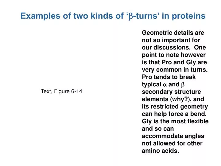
Examples of two kinds of ‘b-turns’ in proteins Geometric details are not so important for our discussions. One point to note however is that Pro and Gly are very common in turns. Pro tends to break typical a and b secondary structure elements (why?), and its restricted geometry can help force a bend. Gly is the most flexible and so can accommodate angles not allowed for other amino acids. Text, Figure 6-14
Different amino acids prefer different types of secondary structures (or turns) • Importance: • Important to take into account if you were engineering different amino acids into a protein • Important for predicting (2°) structure from sequence Text, Table 6-1 Table 6-1
Different classes of protein architecture ‘Globular proteins’: combination of 2° elements into a globular shape, generally with a hydrophobic core, typically soluble, cytosolic, includes most enzymes. Fibrous or filamentous proteins: extended repetition of 2° elements, typically structural, insoluble (e.g. keratin, collagen). Transembrane: restricted combination of 2 ° elements in the lipid bilayer – bundles of a-helices or a b-barrel. Basic elements of secondary structure lipid bilayer
An example of a repetitive structure present in fibrous proteins such as keratin: ‘coiled coils’ Looking down an a-helix: Almost repeats after 7 residues (7*100° = 700°, which is close to but just short of 2 complete turns (720 °). Sometimes a sequence pattern can be seen repeating about every 7 residues (positions a and d being hydrophobic, so that two helices line up side by side). The twisting results from 700° being short of two full turns. Similar (but shorter) coiled-coils (or ‘leucine zippers’) occur also in globular proteins. Figure 6-15a
Another repetitive structure in fibrous proteins: the collagen triple helix • highly repetitive sequence (GXX)n, where X’s are usually proline or hydroxyproline Text, Figure 6-18 Figure 6-18
Fibrous proteins often employ unusual amino acids (and covalent cross-links) hydroxyproline required for collagen synthesis prolyl hydroxylase proline Figure 6-19 Vitamin C deficiency causes scurvy
Fibrous proteins often employ unusual amino acids (and covalent cross-links) Text, Figure 6-19 unusual cross-linking of multiple side chains used to hold together multiple collagen fibrils Figure 6-19
Globular proteins: methods for determining 3D structures at atomic level detail • X-ray crystallography • the dominant method • practically no size limit (e.g. whole ribosome, whole viruses, etc.), as long as crystals can be grown • Multi-dimensional NMR • useful for smaller structures • no crystals required • can give more dynamic information • Electron microscopy • newer instruments are making it possible to approach atomic level detail for special cases (e.g. icosahedral viruses) Figure 6-19
An extreme oversimplification of protein X-ray crystallography X-ray diffraction experiment pure protein an ‘electron density map’, showing the distribution of electron density in 3-dimensions (note that electrons are what scatter X-rays) special computer programs and expertise
In X-ray crystallography, the level of detail you can see is described by the ‘resolution’ Text, Figure 6-23 What the heck? This textbook figure is seriously wrong about what you can see at different levels of resolution. Figure 6-23
NMR structure determination works under different principles • different types of experiments make it possible to establish the secondary structure of different parts of the protein, and to establish distances between different amino acid residues • knowing the distances between groups makes it possible to infer 3D positions for the atoms NMR can give a more dynamic picture of a protein, although uncertainty in positions can be hard to discriminate from dynamics Figure 6-24
Globular protein structure arises from combinations of 2° elements. Some patterns are very common. Text, Figure 6-28 Figure 6-28
Globular protein structures are sometimes classified as all a, all b, or a/b Text, Figure 6-29 Figure 6-29
Even within the same class, tremendous variety is possible. • Two very different antiparallel beta structures are shown. Note the differences in connectivity and overall shape. Figure 6-30a
Larger proteins are generally arranged in multiple ‘domains’. A domain is typically described as an ‘independently folding unit’, which is sometimes hard to define or establish. The protein at the right has two domains. Distinct domains are usually formed by contiguous regions of the primary structure, like an N-terminal domain and a C-terminal domain, but there are exceptions in which the domains are discontinuous. in which case the backbone passes more than once between domains.
Evolutionarily related proteins, whose sequences have diverged very far in evolution, may retain their common structure. This has revealed a tremendous amount of information about protein function, cell biology, and evolution in general. Text: “Thus, it appears that the essential structural and functional elements of proteins, rather than their amino acid residues, are conserved during evolution”. Figure 6-32
Evolutionarily related proteins, whose sequences have diverged very far in evolution, may retain their common structure. This has revealed a tremendous amount of information about protein function, cell biology, and evolution in general. structure of the bacterial ftsZ protein, now understood to be part of a primitive bacterial cytoskeleton. Its amino acid sequence is so divergent from tubulin that no similarity could be detected until the structures were known. structure of the tubulin subunits that assemble to make the eukaryotic microtubule
Quaternary structure (4°): • Types of oligomers (‘oligo’ meaning “few”): • homo-oligomeric (multiple copies of the same subunit) • e.g. a2, a3, a4, etc. (where a refers to the ‘subunit’ identity, not an a helix) • hetero-oligomeric (different protein chains) • e.g. ab, a2b2, a2bg, etc. • sometimes the distinct subunits in a hetero-oligomer are actually similar (e.g. evolutionarily related to each other), making them ‘almost’ homomeric. Hemoglobin (a2b2) is a well known example. Figure 6-33
Quaternary structure (4°): Homo-oligomers (and pseudo homo-olgomers) are nearly always arranged in a symmetric fashion so that every subunit is in essentially the same environment as the other identical ones. This gives rise to two general situations: • essentially linear (or sometimes tubular) filaments or helices • finite symmetric assemblies actin filament a structural protein from a large capsid hemoglobin Figure 6-33
Illustration of the types of symmetry possible for protein assemblies 1 subunit: monomer 2 subunits: dimer 3 subunits: trimer 4 subunits: tetramer 5 subunits: pentamer 6 subunits: hexamer . . . Figure 6-34
Cyclic symmetries Text, Figure 6-34a For larger n, tend to look like a ring (or cycle) of subunits Figure 6-34a
Dihedral symmetries For n>2, tend to look like double-ring structures Figure 6-34b
Cubic symmetries a model of the carboxysome shell • Includes: • some rare enzyme complexes • nearly all protein capsids (i.e. viruses) Figure 6-34c