Download
1 / 28
290 likes | 529 Views
CSULB Radiation Safety Research X-ray Safety Fundamentals. We wish to acknowledge and thank Mr. Adam Weaver, CHP, University of South Florida for the use of this X-Ray training module. It has been modified for use at CSULB. CSULB Radiation Safety Research X-ray Safety Fundamentals.

E N D
CSULB Radiation SafetyResearch X-ray Safety Fundamentals We wish to acknowledge and thank Mr. Adam Weaver, CHP, University of South Florida for the use of this X-Ray training module. It has been modified for use at CSULB.
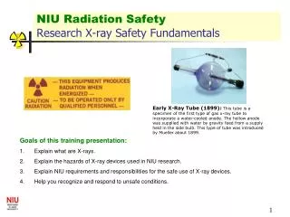
CSULB Radiation SafetyResearch X-ray Safety Fundamentals Early X-Ray Tube (1899): This tube is a specimen of the first type of gas x-ray tube to incorporate a water-cooled anode. The hollow anode was supplied with water by gravity feed from a supply held in the side bulb. This type of tube was introduced by Mueller about 1899. • Goals of this training presentation: • Explain what X-rays are. • Explain the hazards of X-ray devices used at CSULB. • Explain requirements and responsibilities for the safe use of X-ray devices. • Help you recognize and respond to unsafe conditions.
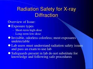
What is radiation? • Radiation is energy in the form of waves or particles. Radiation which is high enough in energy to cause ionization is called ionizing radiation.It includes particles and rays given off by radioactive material and high-voltage equipment. Ionizing radiation includes x-rays, gamma-rays, beta particles, alpha particles, and neutrons. • Without the use of monitoring equipment, humans are not able to "find" ionizing radiation. In contrast to heat, light, odors and noise, humans are not able to see, feel, taste, smell, or hear ionizing radiation.
What are X-rays? • X rays are the ionizing electromagnetic radiation emitted from a highly evacuated high-voltage tube. Inner orbital electrons in the target anode are stimulated to emit x-radiation via bombardment by a stream of electrons from a heated cathode. X-rays, like gamma rays, are penetrating and carry enough energy to ionize atoms in their path. Nearly identical to gamma rays, x-rays require shielding to reduce their intensity and minimize the danger of tissue damage to personnel. Mishaps with x-rays can cause severe radiation burns and deep tissue damage and can lead to various cancers.
X-rays X-rays were discovered in 1895when Wilhelm Conrad Roentgen observed that a screen coated with a barium salt fluoresced when placed near a cathode ray tube. Roentgen concluded that a form of penetrating radiation was being emitted by the cathode ray tube and called the unknown rays, X-rays.
X-ray tube An x-ray tube requires a source of electrons, a means to accelerate the electrons, and a target to stop the high-speed electrons.
X-ray interactions In passing through matter, energy is transferred from the incident x-ray photon to electrons and nuclei in the target material. An electron is ejected from the atom with the subsequent creation of an ion. There are three basic methods in which x-rays interact with matter: photoelectric effect, Compton scattering, and pair production. All these interactions are bad for living cells.
Analytical X-rays • Two X-ray analytical methods are commonly used as research tools at CSULB: Diffraction [XRD] X-ray scattering from crystalline materials yields a “fingerprint” of crystalline structure. Data from the scattered beam is checked against a library of known spectra to identify the material. Fluorescence [XRF] Emission of characteristic "secondary" (or fluorescent) X-rays from a material that has been excited by bombarding with high-energy X-rays.
HAZARDS OF ANALYTICAL X-RAY EQUIPMENT • The primary beam: The primary beam is most hazardous because of the extremely high exposure rates. Exposure rates of 4 x 10⁵ R/min at the port have been reported for ordinary diffraction tubes. 5.0 R is the annual maximum whole body dose allowed to the operator of an x-ray. • Leakage or scatter of the primary beam through cracks in shielding or due to defective equipment: The leakage or scatter of the primary beam through apertures in ill fitting shielding or defective equipment can produce very high intensity beams of possibly small and irregular cross section. • Penetration of the primary beam through the tube housing, shutters or diffraction apparatus: The hazard resulting from penetration of the useful beam through shutters or the x-ray tube housing is slight in well designed equipment. Adequate shielding is easily attained at the energies commonly used for diffraction and florescence analysis. • Diffracted rays: Diffracted beams also tend to be small and irregular in shape. They may be directed at almost any angle with respect to the main beam, and occasionally involve exposure rates of the order of 80 R/h for short periods.
Causes of Radiation Exposure Using ANALYTICAL X-ray • Putting fingers in X-ray beam to change sample • Aligning X-ray beam visually • Modification of shielding • Failure to realize X-rays are emitted from several ports • Failure to read & follow manufacturers X-ray operating instructions Any of these actions could cause an unnecessary exposure and potential serious injury.
Diagnostic X-rays • Two main types of diagnostic X-ray devices: • Radiograph – a picture with film or image is sent direct to computer screen. These are quick ‘shots’. • Fluoroscopic – a real time “moving” inspection on inside functions. These longer exposures yield high doses.
Diagnostic X-rays • Diagnostic radiology is the branch of medicine that involves taking and reading X-rays. The physicians that prescribe the shots and the technologists that operate the machines are specifically trained and licensed to perform these tasks. They also stay current through continuing education. Institutions are always striving to get quality images with the minimum patient exposure.
Industrial X-rays • X-rays are used for non-destructive testing (NDT) and have applications in a wide range of industries. CSULB Engineering classes sometimes use these methods. • NDT uses X-ray beams (or gamma or beta emissions from a radiation source) to inspect products or processes without damaging them. This discipline is called Industrial radiography. • Industrial X-ray machines are used primarily to find defects in castings, structures, and welds, find foreign material in food products, and to inspect luggage at airports, building entries etc. • Electron microscopy equipment generates x-rays inside the apparatus. Factory shielding prevents exposures.
X-Ray Effects • The effects of x-ray exposure depends upon: • Duration/Exposure time - How fast the dose is delivered. • Energy - How much energy was in the x-ray • Low Energy (<50 KeV) - damage only to skin or outer part of body • High Energy - damage to internal organs • Total Dose - The magnitude of the dose
Unsafe conditions • Examples of unsafe conditions which could produce an unwanted radiation dose: Access door interlocks not working, shielding that has been removed/damaged, X-ray “ON” light not lit when unit energized. • IF AN UNSAFE CONDITION ARISES WITH YOUR X-RAY DEVICE: Stop work! Turn power OFF to X-ray @ the cutoff switch Notify X-ray supervisor and Radiation Safety @ 562 985-5623 Post “Do Not Operate” sign at instrument until problem fixed.
RADIATION PROTECTION – TIME The dose of radiation a worker receives is directly proportional to the amount of time spent in a radiation field. Thus, reducing the time by one-half will reduce the radiation dose received by one-half. Operators should always continuously monitor, work quickly and spend as little time as possible next to any high-field X-ray equipment. CSULB presently has no high-field equipment.
Radiation Protection - Distance Radiation exposure decreases rapidly as the distance between the worker and the X-ray device increases. The amount of radiation at a given distance from the source varies inversely with the square of the distance. For example, doubling the distance from an x-ray tube will reduce the dose to one-fourth of its original value, and increasing the distance by a factor of three will reduce the dose to one-ninth of its original value. Maintaining a safe distance, therefore, represents one the simplest and most effective methods for reducing radiation exposure to workers. Using the principle of distance is especially important when working around open beam high-field analytical x-ray equipment.
Radiation Protection - SHIELDING Radiation exposure to personnel can also be reduced by placing material between a worker and the x-ray tube. The energy of the x-ray is reduced by subatomic interactions in the shielding material. Dense materials such as lead, are the most effective shielding, but any metal is useful. Even concrete, water and natural earth are used in shielding. The energy of scattered radiation from x-ray use is often so low that clear plastic is useful. Shielding is often incorporated into the equipment, such as the metal lining surrounding the x-ray tube. It may also consist of permanent barriers such as concrete and lead walls, leaded glass, and plastic movable screens in the case of analytical x-ray equipment.
X-ray device – Open Diffraction This is an OLD open beam X-ray diffraction device. Direct beam injuries are an extreme danger. Newer diffraction X-ray devices for CSULB research must be contained in an fully shielded – interlocked cabinet.
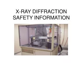
X-ray device – Cabinet Diffraction The X-ray tube, detector and sample are contained in a housing that provides shielding to the user and others in lab. The access doors are interlocked with safety switches and will shut off X-rays when opened. The large viewing area is made possible by effective internal shielding and use of special glass or plastic windows.
X-ray device – Cabinet Diffraction A compact “totally enclosed” research X-ray device. The access door is equipped with a power cutting interlock switch.
X-ray device-Electron Microscope Scanning Electron Microscope (SEM) Transmission Electron Microscope (TEM)
X-ray device – Radiographic Table This is the mobile shield for operator. It is designed to protect operator from scattered X-rays (primarily from patient). This picture X-ray tube in a collimated lead housing. The X-ray beam is pointed down to the table. The table is where the patient is placed and contains a slot for an X-ray film or the newer low-dose digital cassette. This is the control panel. Operator can select X-ray ON (exposure) time in fraction of minutes, the energy of X-ray (in kVp) and current applied (higher current = more X-rays).
State of California Regulations • X-ray devices are regulated by and must be registered with the State of California, Department of Public Health, Radiologic Health Branch. Medical operators must be licensed by this agency and a RHB licensed Supervising physician retained. • Deliberately exposing an individual to an x-ray beam is prohibited unless overseen by a licensed physician in conformance with 17CCR and University policy on human subjects. • Each research x-ray device supervisor shall ensure training of each user and document this training. This training shall consist of Radiation Safety’s introductory training/exam and your machine-specific training. • Each x-ray supervisor shall ensure that x-ray equipment is operated only by persons adequately instructed in safe operating procedures and competent in the safe use of the equipment. Safety rules shall be provided to each operator. A use-log shall be maintained. • The State of California RHB routinely inspects X-ray devices per Title 17 CCR. Registration documents are on file with the RSO.
CSULB requirements for X-ray • If you plan to acquire any X-ray devices YOU MUST get written approval from radiation safety first! 562 985-5623 • CSULB Radiation Safety inspects X-ray devices annually. • X-ray users must be approved and trained by the device Principal Investigator (PI). • Device PI shall ensure that all safety interlocks and shielding are working properly. • Appropriate signs and emergency information shall be posted at the instrument. • X-ray devices shall not be repaired or have the housing removed without prior written approval of the RSO.
Responsibilities of X-ray owners & users • Operate x-ray device only as specified in manufacturers operating instructions and within limits placed by CSULB Radiation Safety. • Notify CSULB Radiation Safety Office of any repairs, modifications, disposal, or relocation of X-ray device.
PERSONNEL MONITORING • Based upon years of monitoring, most analytical X-ray devices at CSULB do not require users to be issued personnel radiation monitoring devices (dosimeters), but they are available. Dosimeters measure and document accrued dose to operators. Calibrated, direct reading meters, are also available from the RSO. • X-ray users should address any radiation safety concerns to CSULB Radiation Safety Office @ 562 985-5623.
Thank you for viewing this X-Ray Training presentation • If you have any questions or comments Please contact CSULB Radiation Safety Office at 562 985-5623 • Jeff Mellon jeff.mellon@csulb.edu • John de la Cuesta jdlc@csulb.edu • Please take the X-ray safety quiz sent to you via email and take it to MIC-001 for grading.