Download
1 / 25
300 likes | 1.06k Views
The Thoracic Cavity. Boundaries of and Structures Within. Body Cavities. Remove frame. Dorsal body cavity Ventral body cavity Thoracic 2 Pleural Mediastinum Divided by Diaphragm Abdominopelvic Abdominal Pelvic. www.newworldencyclopedia.org/entry/Body_cavity.

E N D
The Thoracic Cavity Boundaries of and Structures Within
Body Cavities Remove frame • Dorsal body cavity • Ventral body cavity • Thoracic • 2 Pleural • Mediastinum • Divided by Diaphragm • Abdominopelvic • Abdominal • Pelvic www.newworldencyclopedia.org/entry/Body_cavity
Serous membrane = Serosa • Simple squamous epithelium + areolar connective tissue • 2 Layers • Outer layer = PARIETAL serosa • Inner layer = VISCERAL serosa • Between them = Serous Cavity containing Serous Fluid • Serous fluid is blood filtrate + secretions by 2 layers of membrane • Allows movement of organs with reduced friction • Types of Serous Membranes • Pleural = surrounds lungs • Pericardium = surrounds heart, slightly modified • Peritoneal = surrounds some abdominal organs
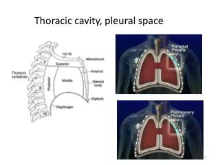
Pleural Cavities • Surround the lungs • Pleural fluid secreted by pleural membranes • Holds layers together • Reduces friction of organs • Benefit of Compartmentalization pg 159
Pleural Cavities • 2 Layers • Visceral pleura (inner) • root of lungs marks transition • external surface of lungs • Parietal pleura (outer) • inner surface of thoracic wall • superior surface of diaphragm • lateral surface of mediastinum pg 161
Pleural Effusion Excess fluid in the pleural cavity More than 20X Usually less than 1 ml of fluid Pneumothorax Air located in pleural space Pleural Abnormalities Pg 238
Divisions of Mediastinum • Superior (to heart) • Contains: thymus, cranial vena cava, trachea, esophagus, nerves • Inferior • Anterior (to heart) • Contains: thymus • Posterior (to heart) • Contains: aorta, esophagus, trachea, bronchi, nerves, caudal vena cava, • Middle • Contains: heart + pericardium pg 177
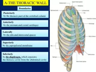
Boundaries of Mediastinum • Lateral • parietal pleura of lungs • Anterior • ventral parietal pleura • Posterior • dorsal parietal pleura • Superior • dome of the neck • Inferior • diaphragmatic pleura pg 159
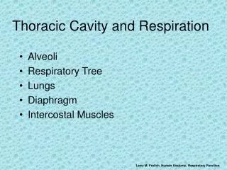
Respiratory Tract • Upper Respiratory Tract • Superior to Larynx • Lower Respiratory Tract • Larynx • Trachea • Primary Bronchi • Secondary Bronchi • Rest of Bronchial Tree • Lungs pg 992 pg 168
Trachea = windpipe • Starts at Larynx and travels through mediastinum • Located Anterior to Esophagus • Trachea terminates into 2 primary bronchi entering lungs • Walls contain 16-20 “C” shaped rings Hyaline Cartilage • Trachealis Muscle (smooth muscle and soft CT) • Layers (deep to superficial) • Mucosa = Ciliated Psuedostratified Epithelium • Submucosa- contains seromucous glands • Adventitia – made of connective tissue, contains cartilage rings pg 966
Bronchial Tree • Primary (main) Bronchi • Bifurcation of trachea • Basically the same structure • Cartilage rings • Posterior to pulmonary vessels • Right is wider, vertical, shorter • Secondary (lobar) Bronchi • Each primary bronchi divides • Same structure as primary bronchi • Right lung has 3, Left has 2 • Tertiary (segmental) Bronchi • Up to 23 divisions pg 168
Bronchial Tree (continued) • Bronchioles • further divisions, < 1 mm diameter • Terminal Bronchioles • further divisions, 0.5 mm diameter • Respiratory Zone • Respiratory Bronchioles • Alveolar Ducts • Alveolar Sacs • Terminal bunches of Alveoli • Respiratory exchange chamber • Among alveoli are blood vessels, nerves, lymphatics www.nlm.nih.gov/.../ency/imagepages/1103.htm
Respiratory Zone (continued) • Lining the Walls of Alveoli • Respiratory Membrane • Type I cells = simple squamous epithelial cells • Basal lamina and fine areolar CT • Covered with capillaries and elastic fibers • Type II cells = cuboidal epithelial cells • Secrete fluid containing surfactant • Dust Cells (macrophages) • Gas exchange • Oxygen into blood • Carbon Dioxide into alveoli
Throughout Bronchial Tree • Psuedostratified columnar changes to simple columnar to simple cuboidal • Cartilage rings replaced by cartilage plates once bronchi enter the lungs • Smooth muscle and Elastic fibers remain important • In Bronchioles • Ciliated mucosa disappears, replaced by macrophages in alveoli • Cartilage disappears • Smooth muscle forms bands around smallest bronchi and bronchioles (not found around alveoli)
LUNGS (continued) • Located in Pleural Compartments • Lateral to Mediastinum • Location • Apex posterior to clavicle • Base lays on Diaphragm • Costal Surface = Ant, Lat, Post surfaces contact ribs • Left Lung = 2 lobes • Upper • Lower • Oblique Fissure • Cardiac Notch • Right Lung = 3 lobes • Upper • Middle • Lower • Oblique fissure • Horizontal fissure pg 168
LUNGS • Hilus- medial indentation • Root of Lung = structures enter each lung • 2 Pulmonary Veins = carries O2-rich blood from each lung to heart • 1 Pulmonary Artery = carries O2-poor blood to each lung • Primary Bronchus • Nerve plexus – Lymph Vessels pg 164
Lung Lobes • Lobes are anatomically + functionally separate • Lung lobes divided into Lobules • Functionally separate • Separated by dense CT • Vary in size • Stroma = lung tissue • Areolar CT • Many elastic fibers pg 178
Esophagus • Esophagus • Pharynx to Stomach • Passes thru diaphragm at esophageal hiatus • Anterior to vertebrae, Posterior to trachea • Layers of Esophagus (deep to superficial) • Mucosa • Stratified squamous epithelium • Lamina propria (loose CT) • Muscularis mucosae • Submucosa • Loose connective tissue • Secretes mucus • Muscularis Externa • Circular/Longitudinal layers • Skeletal m, Mix, then Smooth m • Adventitia • Fibrous CT pg 212
The Diaphragm • Skeletal Muscle • Dome-shaped (relaxed) • Flattens (contracts) • Divides thoracic & abdominopelvic cavities • Attachments • O: Inferior Internal rib cage, Lumbar vertebrae (by crura) • I: Central tendon • Innervated by right + left PHRENIC Nerves pg 136
Action of the Diaphragm • Primary muscle of respiration (involuntary) • Contraction during inspiration • Increases volume of thoracic cavity • Decreases pressure of thoracic cavity • Air moves into lungs (highlow pressure) • Forced contraction (voluntary) • Used for defecation, urination, labor • Decreases volume of abdominal cavity • Increases pressure in abdominal cavity • Pushes on abdominal organs to move contents out pg 136
Thoracic Cavity Capacity is Increased by: • Contraction of diaphragm • Intercostal muscles elevate ribs • Rib elevation causes the sternum to move anteriorly pg 135
Openings of Diaphragm • PosteriorAnterior • Aortic Hiatus • Aorta • Azygos vein • Thoracic duct • Esophageal Hiatus • Esophagus • Vagus nerves • Caval Opening • Inferior Vena Cava • Right Phrenic Nerve pg 157
Superior Vena Cava in Superior mediastinum, right side Receives blood from regions above diaphragm Formed from Rt + Lft Brachiocephalic Veins cranially Azygos Vein empties into it just superior to heart Empties into Right Atrium Inferior Vena Cava in Inferior mediastinum (right side), runs through abdomen Returns blood to heart from regions below diaphragm Formed from Rt + Lft Common Iliac Veins Empties into Right Atrium Widest blood vessel in body Vena Cava
Veins of Thoracic Cavity • Vena Cavae • Azygos Vein • “unpaired” • right side of vertebral bodies (at level of T12) • runs superiorly • empties into Sup. Vena Cava • drains right posterior intercostal veins • Connects to hemiazygos and accessory hemiazygos that drain left side pg 153
Thymus Gland • Lymphatic Organ • 2-lobed w/lobules • Sits on heart and great vessels • Immature lymphocytes mature into T-lymphocytes • Secretes Thymic Hormones: help T-lymphocytes gain immunocompetence • Decreases in size w/age • Functional tissue is replaced with fatty tissue • Contains lobes and lobules • Capsule • Cortex • Medulla pg 206