Download
1 / 38
380 likes | 387 Views
Membrane Dynamics. Composition and Movement within the membrane. Components of the Membrane. Extracellular environment. Carbohydrate. Glycolipid. Phospholipid bilayer. Glycoprotein. Transmembrane (or Integral) protein. Extracellular leaflet. Cytosolic

E N D
Membrane Dynamics Composition and Movement within the membrane
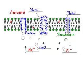
Components of the Membrane Extracellular environment Carbohydrate Glycolipid Phospholipid bilayer Glycoprotein Transmembrane (or Integral) protein Extracellular leaflet Cytosolic Leaflet (varies in content from Extracellular Leaflet) Lipid Anchor Protein Peripheral membrane proteins Cholesterol (found only in animal cells; plants have Phytosterol) Cytosol 4 Major Components of Membranes Phospholipid bilayer (Extracellular and Cytosolic Leaflets have distinct composition) Proteins (Integra/Transmembrane and Peripheral) Carbohydrates (Glycolipids, Glycoproteins) Cholesterol (animals only) or Phytosterols (plants only)
Summary of Membrane Components Phospholipids Bilayer Proteins Carbohydrates Cholesterol or Phytosterol Extracellular Leaflet And Intracellular Leaflet Each has a variety of specific phospholipids, and the two leaflets are also different. • Transmembrane (or Integral) • Extracellular domain (hydrophilic) • Transmembrane segment (hydrophobic) • Intracellular domain (hydrophilic) • Lipid Anchor: covalently bonded to lipid • Peripheral (Extrinsic) non-covalently bound to other membrane proteins or phospholipid heads. • Carbohydrate groups are bonded to some phospholipid heads and some integral proteins • Glycolipids • Glycoproteins • Animals vs. plants respectively • Stabilizes the membrane • Makes more fluid when cold • Makes less fluid when hot
Mosaic Mosaic: something resembling such a picture or decoration in composition, especially in being made up of diverse elements: Cell Membranes are often described as being a Mosaic comprised of different proteins, phospholipids, and carbohydrates. Lateral View Birds-eye View
Fluid Mosaic Model • Fluid Mosaic: Model describing the dynamic structure of membranes • Phospholipid tails are generally unsaturated – membrane is semi-fluid (like oil) • A “mosaic” of integral proteins move freely within the plasma membrane • Components migrate laterally and spin, but don’t flip without an enzyme. • Larger proteins move more slowly Lateral View Birds-eye View Spontaneous lipid movements Lipid movement via flippase Lateral movement Flippase Flip-flop Rotational movement ATP ADP + Pi
Plasma Membrane Unsaturated versus saturated hydrocarbon tails • Factors affecting Fluidity • More saturated tails = less fluid due to improved stacking. • Longer tails = less fluid due to stronger Van der Waals interaction • Low Temperature = less fluid Fluid Viscous Unsaturated hydrocarbon tails Saturated hydrocarbon tails Without Cholesterol • Cholesterolmoderates the fluidity, increasing it at low temperatures and decreasing it at high temperatures(generally it decreases fluidity, unless the organism lives in very cold environments) Fluidity With Cholesterol Temperature
Metabolic Pathways OH HO OH CH2 HO OH H2N CH2 H2N COOH CH2 H2N COOH OH HO OH HO OH H2N OH H2N 1 2 3 4 • Metabolic Pathway: Multiple enzymes act in sequence to produce a final produce • Products become substrate for the next enzyme, etc. • Some metabolic pathways involve several enzymes dissolved in the cytosol BUT… • Many metabolic pathways involve enzymes embedded inside a membrane so that the products of one enzymatic reaction can be passed directly to the next enzyme in the sequence Dopamine β-hydroxylase Dopa Decarboxylase Phenylethanolamine N-methyl transferase Tyrosine Hydroxylase 1 2 3 4 Epinephrine Tyrosine DOPA Dopamine Norepinephrine • Anabolic: Metabolic pathways which build larger molecules via synthesis reactions • Catabolic: Metabolic pathways which break down large molecules via decomposition Note: the pathway above would be neither Anabolic nor Catabolic, just metabolic.
Lipid Rafts CH3 CH3 CH3 CH3 CH3 CH3 CH3 CH3 CH3 CH3 CH3 CH3 CH3 CH3 CH3 CH3 CH3 CH3 CH3 CH3 OH OH OH OH • Lipid Rafts: Localized Region of membranes where components move together within the mosaic • Less Fluid due to • Saturated Lipid tails • Longer fatty acyl tails • Higher amount of cholesterol • Proteins associated with the “Lipid Raft” are kept close together...good for metabolic pathways • Like a pat of butter floating in a pan of cool oil Inside the cell
Cell Membranes • Cell Membrane: any phospholipid Bilayer associated with a cell • Plasma Membrane: the cell membrane that surrounds the outside of a cell • Organelle Membrane: Internal cell membranes that form compartments specialized for carrying out very specialized tasks (only found in eukaryotic cells) Fungal Cell Prokaryotic Cell Animal cell Plant cell
Membrane Transport Dr. Jason R. Mayberry Castle View High School
Diffusion 101 • Concentration: • The amount of solute dissolved in a given amount of solvent (water) • Usually measured in Molarity (M) or milliMolarity (mM) or mg/mL • Concentration Gradient: • Spatial differences in the concentration of a given solute • Diffusion: • Net movement of particles from HIGH to LOW concentration (down its concentration gradient) • The net direction each type of particle will move depends alone on its own concentration gradient, regardless of the overall concentration of the solution. • Equilibrium: • When the concentration of a given type of particle is even throughout a system • Particles still move around, but there is no net movement in any direction. Low Concen-tration High Concen-tration High Concen-tration Low Concen-tration Dynamic Equilibrium Dynamic Equilibrium
Diffusion 101 • Now imagine that a phospholipid bilayer (a membrane) separates two compartments that have a different concentration of solutes. • What will happen? Low Concen-tration High Concen-tration High Concen-tration Low Concen-tration
Plasma Membrane H H O H N C C OH R OH OH O OH Estradiol testosterone CH2OH O HO OH OH H H OH • Semipermeability: Membranes act as a barrier which prevent some molecules from crossing while allowing others to do so • Hydrophobic “Core” of the plasma membrane blocks polar and charged molecules from passing though it • Molecules that are hydrophobic and/or small can pass through the membrane • Transport Proteins (Channel sand Carriers) provide a means for specific particles to move across the membrane. • Speed of movement depends on • How polar/hydrophobic it is • Size of molecule • Size of gradient across membrane • Number of transporters CO2 O2 EtOH Na+ H2O Ion Channel Inside the cell
Permeability of Different Molecules Gases CO2 N2 O2 Very small, uncharged molecules Ethanol High permeability CH3-CH2-OH Water H2O Moderate permeability Urea H2NCONH2 Sugars Polar organic molecules Low permeability C6H12O6 Na+, K+, Mg2+, Ca2+, Ions Cl– Charged polar molecules and macro- molecules Amino acids ATP Proteins Polysaccharides Nucleic acids (DNA and RNA) Very low permeability
Passive Transport • Most solutes exist in different concentrations on opposite sides of the plasma and organelle membranes. • Passive Transport: movement of molecules down their concentration gradient • Simple Diffusion: • Particles to which the membrane are permeable move freely across the membrane as if it weren’t there. • Facilitated Diffusion: • Particles which are impermeable to the membrane must have a Transport Protein in order to diffuse across the membrane (See Slide 11) • Each type of transport protein generally allows only one type of particle to pass
Active Transport • Energy of Concentration • Energy resulting from a concentration gradient, which is used to cause particles to diffuse • The larger the concentration gradient is, the more energy is available to move particles Secondary Active (Cotransport) Chemiosmosis (Passive) Primary Active ATP Synthesis Uniport Symport Antiport • Active Transport: when the energy from another source can be used to increase a molecules concentration gradient across a membrane. • Primary Active Transport: • Protein pumps use ATP energy to move specific particles UP their concentration gradient (making it more concentrated) • ATP energy Energy of Concentration • Secondary Active Transport: • The energy given off by one particle moving DOWN its concentration gradient is used to move a different particle UP its own • i.e. The movement of one particle is secondary to the movement of another. • Symport: two particles move in the SAME direction • Antiport: two particles move in the OPPOSITE direction. High Energy Higher Energy ADP + Pi ADP + Pi ATP ATP Low Energy • Clarifications • Uni- Sym- and Anti-porter • refers only to the number and direction of movement. • Can be used in Facilitated Diffusion or Active Transport (primary or secondary) • Chemiosmosis: Energy of concentration can be captured to make ATP or perform some other task (Passive diffusion)
Transport Proteins: Channels versus Carriers • Channels: open passageways through which molecules flow freely • Only Used in Facilitated Diffusion • May be gated to control when molecules can move • Allows a particle to move in either direction, depending on the direction of its concentration gradient. • Cannot move molecules against their concentration gradient • Carriers: Conformational changes move molecules from one side of the membrane to the other • Can be used in Facilitated Diffusion or Active Transport (primary or secondary) • Movement can be regulated Gate opened Gate closed Uniporter: 1 solute moves in one direction (e.g. primary active) Symporter: 2 solutes move in same direction. Antiporter: 2 solutes move in opposite directions.
Types of Gates on Protein Channels • Channels can regulate the movement (turn it on and off) by opening and closing gates. • Types of Gates • Leaky: always open (No gate/not regulated). • Ligand (Chemically) Gated: open in response to a specific chemical (i.e. a neurotransmitter) • Mechanically gated: open in response to physical deformation of the receptor (i.e. touch) • Voltage Gated: only open in response to specific electrical changes Extracellular Cl- K+ Cl- K+ Cl- K+ Cl- Cl- K+ Cl- Cl- K+ K+ Cl- K+ K+ Cl- Intracellular
Summary of Types of Transport Transport Passive Active Down the Conc. Gradient Up the Conc. Gradient Facilitated Diffusion Primary Secondary Simple Diffusion Uses ATP; Carriers also called Pumps Uses Energy of Concentration; also called Pumps Uses Channels Or Carriers Leaky (ungated) Ligand Gated Mechanically Gated Voltage Gated Photon Gated Chemiosmosis Molecules move directly through the phospholipid bilayer; No Transport Proteins Used. Uniporter n.a. ATP Symporter ATP Cotransporters Antiporter ATP
Concentration Gradients and Transport Maximums • The rate of transport varies with the concentration gradient for all types of transport • Each type of transport varies in unique ways. • Simple diffusion • Always move from high to low • Rate is linearly related to the size of the gradient • Rate is only limited by thermodynamic properties • Facilitated Diffusion • Always moves from high to low • Rate reaches a maximum per the number of transporters • Active Transport • Always moves in one direction (usually low to high) • Rate declines as the concentration gradient increases • Rate Reaches a Maximum per the number of transporters For a small nonpolar molecule (e.g. O2) Fast In For semipermeable molecules (e.g. H20) 100 0 50 0 Rate of Transport 50 50 50 Fast Out Extracellular Solute Concentration Relative to Intracellular Gradient is large – gradient is zero Gradient is large + small equal large Simple Diffusion Active Transport (Exporter) Facilitated Diffusion Active Transport (Importer)
Dynamics of Transport (Simplified Graph) Rate of Solute Transport Across the Plasma Membrane • Graphs showing Transport Dynamics are often simplified to illustrate the key aspects of each type of transport. • Simple: • Linear with Size of Gradient • Facilitated: • Limited by the Number of Transporters • Active: • Limited by Number of transporters when gradient is small • decreases to zero when working against a large gradient. Rate of Transport Size of Concentration Gradient Simple Diffusion(moving down the gradient) Facilitated Diffusion(moving down the gradient) Active Transport(moving against the gradient)
Sodium (Na) – Potassium (K) Pump (a Primary Active Antiporter) Extracellularenvironment Positively (+)Charged 2 K+ E2 E1 E2 Conformation E1 Conformation Pi P ADP + ATP Intracellularenvironment 3 Na+ Negatively (–)Charged • Found in all Animal Cell Membranes • Uses ATP to move 3Na+ out and 2K+ in • Establishes a Concentration gradient of Na+ and K+ • Net movement of + out results in a small electrochemical gradient across the cell • Electrical: + out/- in • Chemical: Na high concentration out • Chemical: K high concentration in • Energy stored in the electrochemical gradient is used to fuel many cellular processes
Proton Pump (a Primary Active Uniporter)andSucrose – H+Cotransporter(a Secondary Active Symporter) • Proton Pumps and Sucrose Cotransporters are found in plant cell membranes. • Coupling of the primary active transporter with symportenables plants to concentrate sucrose molecules where needed. ATP H H Proton pump H H H H H H Sucrose-Hcotransporter Diffusion of H Sucrose Sucrose
Movement of Large Particles Across a Membrane Exocytosis and Endocytosis
Moving LARGER Particles across the Membrane Exocytosis: Movement of materials out of the cell by fusing a vesicle with the plasma membrane SNARE proteins Endocytosis: • Pinocytosis: “Cell Drinking”Brings a small volume of extracellular fluid and the solutes suspended in it into the cell; non Specific • Receptor Mediated EndocytosisReceptors on the cell surface bind specific molecules and are then Endocytosed with the bound molecule. • Phagocytosis: “Cell Eating”Large objects brought into the cell by wrapping the plasma membrane around it then fusing the extensions together.
Exocytosis and Endocytosis Exocytosis Receptor-Mediated Endocytosis Pinocytosis (Cell drinking) Phagocytosis (Cell eating) EXTRACELLULARFLUID Solutes Pseudopodium Movement of materials out of the cell by fusing a vesicle with the plasma membrane Receptor Ligand Plasmamembrane Coat proteins Coatedpit “Food” orother particle Receptors on the cell surface bind specific molecules and are then Endocytosed with the bound molecule. Proteins lining a portion of the plasma side of the membrane 1) causing it to pinch inward and 2) allowing identification of the contents Coatedvesicle Brings a small volume of extracellular fluid and the solutes suspended in it into the cell; non Specific Vesicle Large objects brought into the cell by wrapping the plasma membrane around it then fusing the extensions together. Foodvacuole
Movement of Water • ASSUME BARRIER IS PERMEABLE TO SOLUTES • Diffusion: Each solute will move relative to its own concentration (Molarity), from high to low Molarity. • ASSUME BARRIER IS IMPERMEABLE TO SOLUTES • Osmosis: Water moves relative to the total solute concentration (Osmolarity), from low to high osmolarity. (i.e. water is “color” blind) Na+ Cl- Glucose Na+ Cl- Glucose Permeable to all solutes and water Permeable only to water 5mM 1mM 5mM 1mM 2mM 7mM 2mM 7mM 3mM 4mM 3mM 4mM Water 10mOsM 12mOsM
The Movement of Water: Osmosis • Osmolarity • Measures the number of particles per liter dissolved in water (Moles of particles/liter) • Ionic compounds produce two different particles when dissolved • Covalently bonded compounds only produce one particle • Osmosis: • Movement of water across a membrane • Water doesn’t care what type of particles are on either side of the membrane only the number of particles • Water will always move from LOW to HIGH Solute Concentration (i.e. osmolarity) • Osmotic Pressure • The pressure exerted by a solvent (such as water) when passing through a membrane. • Can cause water to “defy gravity” • Pressure is equal to the force needed to prevent (or reverse) its movement. A force equal to the osmotic pressure would make the water levels equal. 1Mole of NaCl 1Mole of Glucose 2 Osmoles 1 Osmole 1 Molar 1 Molar Semipermeable membrane(impermeable to Na+, Cl-, and Glucose) 2 Liters of Solution Membrane Permeable to solutes
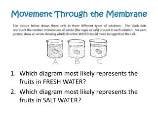
Lowerconcentrationof solute (sugar) Higher concentrationof solute Same concentrationof solute Sugarmolecule Figure 7.14 H2O Selectivelypermeablemembrane Osmosis
Which way will each solute diffuse? Which way will water move? Na+ Cl- Glucose Permeable only to Na+ and water Initial Concentrations Concentrations after Na+ Moves 5mM 1mM 3mM 3mM 2mM 7mM 2mM 7mM Water 5mM 4mM 5mM 4mM 12mOsM 12mOsM 10mOsM 14mOsM + Osmotic Pressure • Na+ will move down its concentration gradient • When Na+ moves it disrupts the OsM balance. • Water will follow Na+ • Pressure (Osmotic Pressure) will increase on the right side • Because membranes are generally semi-permeable to water, it will normally follow solutes when they move, though perhaps more slowly.
Tonicity • Tonicity: • A measure of the ability of a solution to cause water to flow across a membrane into our out of a cell • Compares relative Osmolarity across a membrane 5mOsm 10mOsm 15mOsm 10mOsm 10mOsm 10mOsm Hypotonic: solution has lower Osmolarity than the cell Isotonic: Solution has the same Osmolarity as the cell Hypertonic: The solution has higher Osmolarity than the cell
Osmosis in Animals and Plant Cells Osmosis in animal cells Osmosis in plant cells Isotonic Isotonic Flaccid: plasma membrane is touching the cell wall, but is not exerting pressure on it. Animal cell has normal shape Extracellular Fluid is Hypertonic Extracellular Fluid is Hypotonic Extracellular Fluid is Hypertonic Extracellular Fluid is Hypotonic Crenation: water loss from the cell causes the cell to shrivle Lysis: Pressure from water gain causes the cell to burst open Turgid: Cell Wall Prevents Expansion; Osmotic pressure on teh cell wall makes the plant stiff (Normal) Plasmolysis leads to Wilting and cell death
More of the same (Figure 7.15) Isotonicsolution Hypertonicsolution Hypotonicsolution Animal cell H2O H2O H2O H2O Shriveled (Crenated) Lysed Normal Cell wall H2O H2O H2O H2O Plant cell Turgid (normal) Flaccid Plasmolyzed
Diffusion of Water • Simple Diffusion: water moves somewhat slowly in and out of all cells by diffusion through the plasma membrane • Facilitated Diffusion: some cells (root hairs, red blood cells, kidney tubules) have a channel named aquaporinwhich allows water to move more quickly • WATER IS NEVER MOVED BY ACTIVE TRANSPORT!!! Facilitated diffusion of Water Aquaporin Simple diffusion of Water