Download
1 / 25
341 likes | 784 Views
Structure and function of hemoglobin. GIT | 1 Lecture | Dr. Usman Ghani. Hemoglobin (Hb). A hemeprotein found only in red blood cells Oxygen transport function Contains heme as prosthetic group Heme reversibly binds to oxygen. The heme group.

E N D
Structure and function of hemoglobin GIT | 1 Lecture | Dr. Usman Ghani
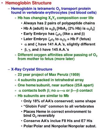
Hemoglobin (Hb) A hemeprotein found only in red blood cells Oxygen transport function Contains heme as prosthetic group Heme reversibly binds to oxygen
The heme group • A complex of protoporphyrin IX and ferrous iron (Fe2+) • Fe2+ is present in the center of the heme • Fe2+ binds to four nitrogen atoms of the porphyrin ring • Forms two additional bonds with: • Histidine residue of globin chain • Oxygen
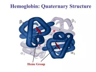
Hemoglobin A (HbA) • Major Hb in adults • Composed of four polypetide chains: • Two α and two β chains • Contains two dimers of ab subunits • Held together by non-covalent interactions • Each chain is a subunit with a heme group in the center that carries oxygen • A Hb molecule contains 4 heme groups and carries 4 moelcules of O2
T-form of Hb The deoxy form of Hb Taut form The movement of dimers is constrained Low-oxygen-affinity form
R-form of Hb The oxygenated form of Hb Relaxed form The dimers have more freedom of movement High-oxygen-affinity form
Hemoglobin function • Carries oxygen from the lungs to tissues • Carries carbon dioxide from tissues back to the lungs • Normal level (g/dL): • Males: 14-16 • Females: 13-15
Factors affecting oxygen binding • Three allosteric effectors: • pO2 (partial oxygen pressure) • pH of the environment • pCO2 (partial carbon dioxide pressure) • Availability of 2,3-bisphosphoglycerate
Oxygen Dissociation Curve The curve is sigmoidal Indicates cooperation of subunits in O2 binding Binding of O2 to one heme group increases O2 affinity of others Heme-heme interaction
P50 • Indicates affinity of Hb to O2 • P50(mm Hg): the pressure at which Hb is 50% saturated with O2 • High affinity slow unloading of O2 • Low affinity fast unloading of O2 • Lung pO2 is 100 mm Hb saturation 100% • Tissue pO2 is 40 mm Hb saturation reduces • Hence O2 is delivered to tissues
The Bohr effect • Effect of pH and pCO2 on: • Oxygenation of Hb in the lungs • Deoxygenation in tissues • Tissues have lower pH (acidic) than lungs • Due to proton generation: • CO2 + H20 HCO3- + H+ • Protons reduce O2 affinity of Hb
The Bohr Effect Causing easier O2 release into the tissues The free Hb binds to two protons Protons are released and react with HCO3– to form CO2 gas The proton-poor Hb now has greater affinity for O2 (in lungs) The Bohr effect removes insoluble CO2 from blood stream Produces soluble bicarbonate
Availability of 2,3 bisphosphoglycerate • At high altitudes: • -RBC number increases • Hb conc. increases • BPG increases Binds to deoxy-hb and stabilizes the T-form When oxygen binds to Hb, BPG is released
High altitude and O2 affinity • In hypoxia and high altitude • 2,3 BPG levels rise • This decreases O2 affinity of Hb • Thus increases O2 delivery to tissues
High O2 affinity High O2 affinity is due to: • Alkalosis • High levels of Hb F • Multiple transfusion of 2,3 DPG-depleted blood
Fetal Hemoglobin (HbF) Major hemoglobin found in the fetus and newborn Tetramer with two a and two g chains Higher affinity for O2 than HBA Transfers O2 from maternal to fetal circulation across placenta
HbA2 Appears ~12 weeks after birth Constitutes ~2% of total Hb Composed of two a and two d globin chains
HbA1c HbA undergoes non-enzymatic glycosylation Glycosylation depends on plasma glucose levels HbA1c levels are high in patients with diabetes mellitus
Abnormal Hbs Unable to transport O2 due to abnormal structure Carboxy-Hb: CO replaces O2 and binds 200X tighter than O2 (in smokers) Met-Hb: Contains oxidized Fe3+ (~2%) that cannot carry O2 Sulf-HB: Forms due to high sulfur levels in blood (irreversible reaction)