Download
1 / 30
320 likes | 917 Views
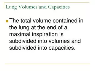
Pulmonary Volumes and Capacities ( Spirogram ). DR QAZI IMTIAZ RASOOL. OBJECTIVES. Describe normal spirogram , labeling, defining and mentioning the normal values of lung volumes and capacities.

E N D
Pulmonary Volumes and Capacities(Spirogram) DR QAZI IMTIAZ RASOOL
OBJECTIVES • Describe normal spirogram, labeling, defining and mentioning the normal values of lung volumes and capacities. • Discuss and identify volume and capacities that cannot be measured by spirometry and discuss their significance and factors affecting them. • Define dead space, identify its different types and mention its significance. • Describe the changes in these volumes and capacities in obstructive and restrictive pulmonary diseases.
Pulmonary function tests are group of procedures that are designed to measure (evaluate) the functions of the lung. • Examples include: • Spirometry. • Lung volumes by helium dilution or body plethysmography. • Blood gases. • Exercise tests. • Diffusion capacity. • Bronchial challenge testing. • Pulse oximetry.
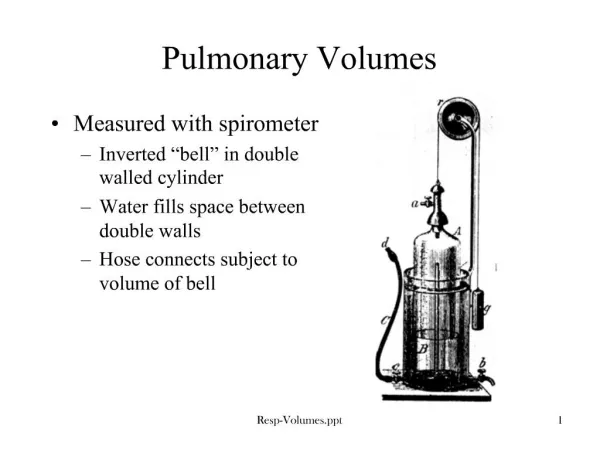
SpirometryDescribe the measurement of lung volume Basic tools to know respiratory status. • In patients with pulmonary disease, • As a first diagnostic test employed In pre-operative evaluation, 3.In managing patients with pulmonary disease, 4. In quantifying pulmonary disability. 5.In evaluation of allergic status for drug therapy 6.As epidemiological survey to know normal values 7.To assess impact of an occupational exposure
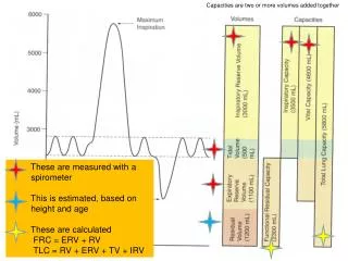
Spirogram Volumes Capacities Vital Capacity V.C Total Lung Capacity T.L.C Function Residual Capacity F.R.C Inspiratory Capacity I.C • Tidal Volume (Minute)T.V • Residual Volume R.V • Inspiratory Reserve Volume I.R.V • Expiratory Reserve Volume E.R.V
-Lung volume can be measured by;- 1. Changes of the lung volume during one breathing at static conditions, called static/ Primarylung volume Or 2. Same / unit time called dynamic/ secondary lung volume
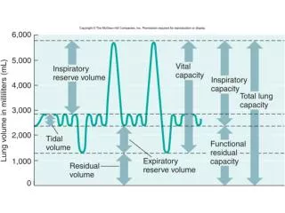
Tidal volume = volume of air that can be inhaled or expelled during normal quiet breathing = 500 ml. Inspiratory reserve volume = Maximum volume of air that can be inspired by forced inspiration after normal inspiration = 3000 ml. Expiratory reserve volume = Maximum volume of air that can be expired by forced expiration after normal expiration = 1000 ml. Residual volume = volume of air that remains in the lung after forced expiration = 1200 ml. 1. Can be expelled from the lung unless the thoraxic cavity is opened as in pneumothorax 2. Physiological significance: aerates blood in between respiratory cycle 3. Clinical significance: RV / TLC should by 25-30% . If more obstruction of airways is diagnosed as in bronchial asthma 4. Not measured by spirometer. It is mesured by helium dilution
Inspiratory capacity= the maximum volume of air that can be inspired by forced inspiration after normal expiration = TV + IRV = 3500 ml. Functional residual capacity = Volume of air that remains in the lung after normal expiration = RV + ERV = 1200 + 1000 = 2200 ml Not measured by spirometer. Total lung capacity= volume of air that is contained in the lung after forced inspiration = IRV + TV+ ERV + RV = 5700 ml. Not measured by spirometer.
2. Indirect SpirometryGas dilutionBody plethysmography Gas dilution techniques • All operate on a principle SIMILAR to Boyle’s Law (P1 V1 = P2 V2), C1 V1 = C2 V2 NOTE;- 1. Can only measure lung volumes in communication with conducting airways 2. Obstruction or bullous disease can have trapped, noncommunicating air within the lungs (FRC may be measured is less than its actual volume)
Static lung volumes & capacities, • RV, FRC, & TLC cannot be determined with • direct spirometry. i.e, Closed-Circuit Helium dilution method for RV, FRC, TLC Usually first FRC is calculated 1. FRC= ([He]i/([He]f-1)Vi • [He]i=initial concentration of helium in spirometer • [He]f=final concentration of helium in spirometer • Vi=initial volume of air in bell of spirometer 2. RV = FRC- ERV 3. TLC= RV + VC
Vital capacity: the maximum volume of air that can be expired by forced expiration after forced inspiration = IRV + TV + ERV = 4500 ml. It is the second best pulmonary function test. Factors affecting VC: Posture Abdominal content Respiratory muscles Thoracic wall Resistance to airflow Lung elasticity Pulmonary blood volume
Peak Expiratory Flow Rate (PEFR) • Measuring maximal velocity of expired air after forceful inspiration. • Using peak flow meter. • Values for age, sex, height • Alternative for FEV/FVC. • Reduced in COPD. • Of special importance in asthma for rapid follow up of treatment efficacy. PEFR (L/min) = [Height (cm) - 80] x 5
Maximal Voluntary Ventilation (MVV) or Maximum Breathing Capacity (MBC): It is the maximum volume of air that can be breathed in and blownout per minute using the fastest and deepest respiratory effort as ever. Procedure :Breathing as rapid & as deep as possible for 15 sec then multiply the result x 4. Equals 80-160 L/min in ♂ and 60-120 L/min in ♀ It is the best pulmonary function test. Breathing Reserve = MVV-MPV Equals100 - 8 = 92 L/min Dyspnoea Index = Breathing Reserve/MBC Normally = 90% If < 60% , dyspnoea is diagnosed.
Dead Space The space that contains the volume of air that dose not undergo gas exchange with the blood in the pulmonary capillaries. Dead space is filled with inhaled air at the end of inspiration & with alveolar air at the end of expiration. Volume of air in dead space = 150 ml. Types of Dead space: Anatomical; conducting zone of respiratory tract. Alveolar DS; non-perfused alveoli Physiological DS = anatomical D.S + alveolar DS In normal subjects: anatomical dead space = physiological dead space as there is no non-functional alveoli. In diseased subjects: Physiological dead space > anatomical dead space.
Obstructed Airflowlimitation of expiratory airflow so that airways cannot empty as rapidly compared to normal • Narrowing of the airways due to bronchial smooth muscle contraction i.e. Asthma • Narrowing of the airways due to inflammation and swelling of bronchial mucosa and the hypertrophy and hyperplasia of bronchial glands i.e, bronchitis • Material inside the bronchial passageways physically obstructing the flow of air i.e excessive mucus plugging, inhalation of foreign objects or the presence of pushing and invasive tumors • Destruction of lung tissue with the loss of elasticity and hence the loss of the external support of the airways i.e. Emphysema • External compression of the airways i.e.Tumors, trauma
Restricted AirflowCharacterized by reduced lung volumes/decreased lung compliance A. Intrinsic Restrictive Lung Disorders 1. Sarcoidosis 2. Tuberculosis 3. Pnuemonectomy (loss of lung) 4. Pneumonia B. Extrinsic Restrictive Lung Disorders 1. Scoliosis, Kyphosis 2. AnkylosingSpondylitis 3. Pleural Effusion 4. Pregnancy 5. Gross Obesity 6. Tumors 7. Ascites 8. Pain on inspiration - pleurisy, rib fractures C. Neuromuscular Restrictive Lung Disorders 1. Generalized Weakness – malnutrition 2. Paralysis of the diaphragm3. Myasthenia Gravis - in which the nerve impulses fail to induce muscular contraction. 4. Muscular Dystrophy 5. Poliomyelitis 6. Amyotrophic Lateral Sclerosis
Flow/Volume Loops in Obstruction and Restriction • ↓VC, normal shape reduced. • FVC is normal. • concave, scooped appearing • VC is normal.FEV1 is reduced. General rule: when flow is ↓→ lesion is obstructive When volume is↓→it is restrictive
How is a flow-volume loop helpful clinically? • Helpful in evaluation of air flow limitation on inspiration and expiration • In addition to obstructive and restrictive patterns, flow-volume loops can show provide information on upper airway obstruction: • Fixed obstruction: constant airflow limitation on inspiration and expiration—such as in tumor, tracheal stenosis • Variable extrathoracic obstruction: limitation of inspiratory flow, flattened inspiratory loop—such as in vocal cord dysfunction • Variable intrathoracic obstruction: flattening of expiratory limb; as in malignancy or tracheomalacia
Spirometry Interpretation: What do the numbers mean? FEV1 >75% 60%-75% 50-59% <49% FVC 80-120% Normal 70-79% Mild reduction 50%-69% Moderate <50% Severe
Classification of Ventilative Function Disorder obstruction restriction mixed FEV1/FVC N or MVV or N VC N or RV uncertain TLC N or N or uncertain
Effects of Aging • VC and MVV ↓ • RV and DS ↑ • Ability to remove mucus from respiratory passageways ↓ • Gas exchange across respiratory membrane ↓
Critical Thinking In the advanced stages of pulmonary emphysema, the FRC and the RV are increased; in addition the VC is often decreased. Why do these changes occur ?