Download
1 / 90
1.26k likes | 3.11k Views
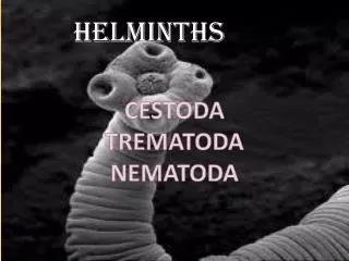
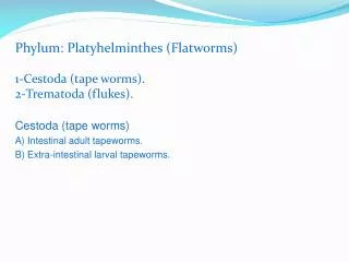
The Cestodes (tape worm). Nematode trematode. common features of Class Cestoda. 1. Adult worm is flattened ribbon-like 2. Body wall: Tegument and subtegument; and no body cavity. body wall construction :. substantia corticalis :

E N D
The Cestodes (tape worm)
common features of Class Cestoda 1. Adult worm is flattened ribbon-like 2. Body wall: Tegument and subtegument; and no body cavity.
body wall construction: • substantia corticalis: • microvilli、cytomere、basal membrane • ①scratch intestinal mucosa • ②conduce(to)attachment • ③expand surface areaof absorption ④possess sensory function • ⑤to resist digestion subcutaneous stratum: annuliform muscle、vertical muscle、oblique muscle help the proglottid Slipping and falling off
3.The body is composed of a head, neck and segmented strobilus (链体). The head has suckers, rostellum (顶突) and hooklets or sucking grooves. The neck is the budding zone from which segments are formed. The strobilus consists of immature, mature and pregnant proglottides.
Strobila (链体): (1)Scolex (头节) - equipped with organs of attachment: suckers, hooks, grooves. (2)Neck (颈部)- germinal portion (3) Proglottids (节片) - immature, mature, gravid(孕节).
4. They are hermaphroditic. There is a set of female and male reproductive organs in every mature proglottid.
5. Digestive tract is absent. Nutrition is absorbed by villi of body surface. Nematode trematode
6. They are biohelminths. Intermediate hosts are indispensable. 7. All adult worms parasitize digestive tracts of mammals.
8.The developing stages in intermediate hosts are called metacestode (中绦期), such as cysticercus (囊尾蚴), hydatid cyst (棘球蚴), cysticercoid (似囊尾蚴), procercoid (原尾蚴), plerocercoid(裂头蚴).
PHYSIOLOGY AND PATHOGENESIS: • 1. Surface absorption capabilities. • 2. Highly developed reproductive • functions. • 3. Anaerobic metabolism • (无氧代谢).
PHYSIOLOGY AND PATHOGENESIS: (2) • 4. No external environment • development. • 5. All species are parasitic. • 6. Pathogenic stage may be • adult or the larvae.
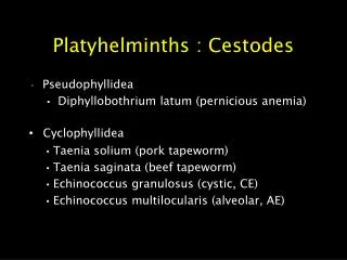
Tapeworms are classified into two orders: (1).Cyclophyllidea (圆叶目):The head is spherical with suckers, hooklets. The uterus has no opening. One intermediate host is required. The eggs contain an oncosphere (六钩蚴). They are medically important, such as Taenia solium (链状带绦虫), Taenia saginata (肥胖带绦虫) and Echinococcus granulosus (细粒棘球绦虫).
(2).Pseudophyllidea (假叶目):The head is spear-like with sucking grooves. The uterus has an opening. Two or more intermediate hosts are required. The eggs contain a coracidium(钩球蚴) and have to get into water to develop. Human being occasionally get infection. This worms include Spirometra mansoni(曼氏迭宫绦虫) and Diphyllobothrium latum(阔节裂头绦虫).
Pseudophyllidea (假叶目): Cyclophyllidea (圆叶目)
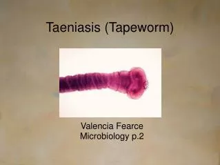
Taenia solium(pork tapeworm) Adult worms live in human small intestine causing taeniasis (绦虫病). The larval stage (Cysticercus cellulose 猪囊尾蚴) lives in pig or human tissues causing human cysticercosis(囊尾蚴病或囊虫病).
GENERAL INTRODUCTION: 1. Worldwide distribution. 2.Large tapeworm. 3. Taenia solium: Laval infection cause serious clinical disease --- CYSTICERCOSIS.
Adult: • Taenia solium (pork tapeworm)
Adult of the Pork Tapeworm averages 2 – 4 meters long.
I. Morphology 1. Adultis flattened ribbon-like, creamy white in color, measures about 2-4 m and has 700-1000 proglottides. scolex : global, 1mm. With 4suckers, 1rostellum(顶突) and 25-50 hooklets arranged in a double crown It consists of neck: it’s the narrowest part of the body and budding zone containing germinative tissue immature proglottides:width>length strobila mature proglottides: width=length gravid proglottides: width<length
Immature proglottides are transverse rectangle, located in the anterior part of the body and inner organs are developing. Mature proglottides are square in shape and located in the mid part of the body and have 150-200 testes, a centrally straight uterus and 3 lobes of ovary . Pregnant(gravid) proglottides are longitudinal rectangle, located in the posterior part of the body and contain a branched uterus filled with eggs. The number of main branches on each side of the uterus stem is 7-13.
India Ink Technique • Note : less than 13 lateral uterine branches (one side).In this species the uterus of the gravid proglottid has between 7 and 13 lateral branches on each side.
2. Egg, The eggs of Taenia saginata and T. solium are indistinguishable morphologically. The eggs are spherical, diameter 31 to 43 µm, with a thick radially striated brown embryophore (胚膜). Inside each is an oncosphere (六钩蚴) with 6 hooklets. t
3.Cysticercus cellulose. It is a semitransparent and elliptic bladder, like a white pomegranate seed (石榴籽)about 0.6-1cm. There is fluid and a white scolex with 4 suckers and hooklets in the bladder.
Under stimulation of bile The scolex invaginates in the bladder The scolex evaginates
II. Life cycle 1. final host: man, 2. Intermediate host: swine (or man), 3. Infective stage: cysticercus and egg, 4. Infective mode: eating raw bean-pork or egg 5. Site of inhabitation: adult in small intestine; cysticercus in tissues, 6. Infective mode of cysticercosis: endogenous, exogenous auto-infection and foreign source; 7. Life span: more than 25 years; cysticercus can survives 5-6 years in human body.
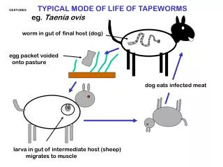
Life Cycle of Taenia solium Attach to intestinal mucosa scolex evaginates adults grivad proglottides develop into fall off duodenum In human small intestine and are 2-3 months discharged in feces man eats cysticercus in raw pork ───────────────────────────── swine ingests eggs In intermediate hosts man gets infection 60-70days develop into cysticercus onchosphere hatch in all parts of the body larval migration in duodenum penetrate intestinal wall into blood stream
III. Pathogenesis and Clinical Manifestations • 1. Taeniasis: It is caused by the adult residing in small intestine of the man. The adult irritates the small intestine causing discomforts, such as abdominal pain, anorexia, chronic indigestion, diarrhea, emaciation and etc. The patient is usually no obvious symptom, only complaining passing proglottides.
2. Cysticercosis: It is caused by the cysticerci living in human tissues. The manifestations vary with the number of cysticerci and the tissues and organs involved. Cysticercosis is divided into four types.
(1) Subcutaneous type: The subcutaneous nodules are usually found in head, limbs, neck, abdomen and back. They are movable and painless.
(3) Ocular type: The cysticercus is usually found in the vitreous body or subretina. Visual disturbance often occurs. The died body of worm may provokes local inflammation causing blindness.
The symptoms are related to the site of infection. The patients may manifest headache, nausea, vomiting, epilepsy (癫痫), paralysis (瘫痪), weakness in limbs, diplopia(复视), dizziness, mental disorder. Epilepsy is the most frequent symptoms of brain cysticercosis. (4) Brain type:
IV. Diagnosis 1.Taeniasis: Confirmative diagnosis of taeniasis is made by finding gravid proglottides or egg in stool. (1) direct fecal smear (2) brine floatation technique **(3) cellophane-tape technique
the examination of a transparent plastic adhesive tape previously applied to the perianal region
Scotch-tape smear Showing some eggs of Taenia spp. and an egg of E.vermicularis.
2. For cysticercosis (1) Specific diagnosis is difficult to establish, the history and adult worm infection attribute to strong suspicion. (2) Biopsy to subcutaneous lesions. (3) computerized axial tomography
(CT) or (MRI). are used for the diagnosis of brain type and ophthalmoscope examination is used for ocular form. 3.Immunological tests are for reference only. IHA, ELISA, Dot-ELISA McAb-CAg.
V. Treatment and prevention 1. Treatment of Taeniasis: (1) Chinese herb medicine: pumpkin seed and areca nut (槟榔) . The recognition of a scolex in the patient’s stool after the application of taenifuge is important. When the entire worm has been expelled, the therapy is successful, otherwise the strobila regrow. (2) Praziquantel may be used.