Download
1 / 24
260 likes | 300 Views
Nerve supply and blood supply of back of thigh. Dr.Qudusia Sultana. OBJECTIVES. By the end of the lecture, the students should be able to: Describe the anatomy (origin, course,Relation & distribution) of the sciatic nerve in thigh. List the branches of the sciatic nerve in thigh.

E N D
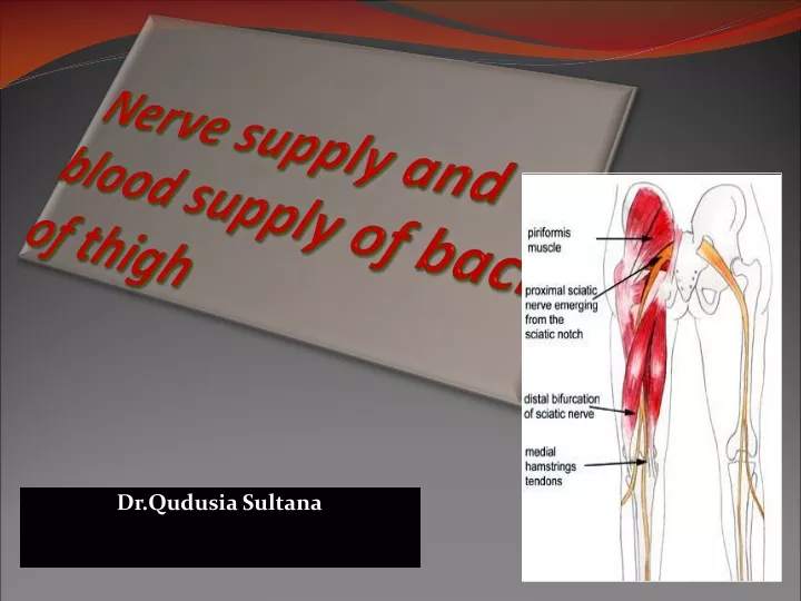
Nerve supply and blood supply of back of thigh Dr.Qudusia Sultana
OBJECTIVES • By the end of the lecture, the students should be able to: • Describe the anatomy (origin, course,Relation & distribution) of the sciatic nerve in thigh. • List the branches of the sciatic nerve in thigh. • Describe briefly the main motor and sensory manifestations in case of injury of the sciatic nerve or its main branches.
The sciatic nerve • The sciatic nerve is a major nerve of the lower limb. • It is a thick flat band, approximately 2cm wide • The largest nerve in the body. • It is composed of 2 parts: • Tibial and common peroneal nerves. • Ventral divisions of anterior primary rami of L4, L5; • S1, S2, S3 compose the tibial part. • Dorsal divisions of anterior primary rami of L4, L5; S1, • S2 compose the common peroneal part.
Origin • The sciatic nerve originates from anterior primary rami of Lumbosacral plexus with root value of L4, L5, S1, S2 and S3
Origin • Tibial division Orginates from ventral division of anterior primary rami of L4,L5,S1,S2,S3. • Common Peroneal division Originates fromDorsal division of anterior primary rami of L4,L5,S1,S2
Note • the sciatic nerve can be described as two individual nerves bundled together in the same connective tissue sheath – the tibial and common peroneal nerves. • These usually separate at the apex of the popliteal fossa, however in approximately • 12% of people they separate as they leave the pelvis.
Site • Formation: • On the posterior pelvic wall. • In front of Piriformis muscle. 4 5
Course • It leaves the pelvis through greater sciatic foramen, below the piriformis & passes in the gluteal region (between ischial tuberosity & greater trochanter) then to posterior compartment of thigh
Relations • DEEP RELATIONS (BED OF THE SCIATIC NERVE) • From above downward the sciatic nerve is related to: -Body of ischium (posterior surface). –Tendon of obturatorinternus –Gemellus superior –Gemellus inferior muscles. –Quadratus femoris. –Adductor magnus.
Relations SUPERFICIAL RELATIONS • From above downward, the sciatic nerve is related to: –Gluteus maximus (in the gluteal region). –Long head of biceps femoris (in the thigh). –The sciatic nerve becomes superficial only in the angle between the gluteus maximus and long head of biceps femoris.
Branches of Sciatic Nerve • Articular branches to the hip joint originate in the gluteal region.
Branches of Sciatic Nerve • 1. Muscular: • To Hamstrings: (flexors of knee & extensors of the hip). Through tibial part to: • Hamstring part of Adductor Magnus. • Long head of Biceps Femoris. • Semitendinosus. • Semimembranosus. Through common peroneal nerve. 1. The short head of biceps
Branches of Sciatic Nerve • 2. Cutaneous: • To all leg & foot EXCEPT: Areas supplied by the saphenous nerve (branch of femoral nerve).
Note • All the muscular branches of the sciatic nerve originate from the medial side with the exception of nerve to short head of biceps femoris, which originates from the lateral side. • Thus, the side lateral to the sciatic nerve is safe side and the side medial to its dangerous side/unsafe side.
CAUSES OF SCIATIC NERVE INJURY • The sciatic nerve is most frequently injured by…? I- Badly placed intramuscular injections in the gluteal region. • To avoid this, injections should be done into the gluteus maximus or medius into the upper outer quadrant of buttock
CAUSES OF SCIATIC NERVE INJURY II-Posterior dislocation of the hip joint
Motor loss • MOTOR EFFECT: • Marked wasting of the muscles below the knee. • Weak flexion of the knee (sartorius & gracilis are intact). • Weak extension of hip (gluteus maximus is intact). • All the muscles below the knee are paralyzed, and the weight of the foot causes it to assume the plantar-flexed position, or Foot Drop. • (Stamping gait).
Sensory Loss • Sensation is lost below the knee,except for a narrow area down the medial side of the lower part of the leg (blue)and along the medial border of the foot as far as the ball of the big toe, which is supplied by the saphenous nerve (femoral nerve).
SCIATICA • Sciatica describes the condition in which patients have pain along the sensory distribution of the sciatic nerve. • Thus the pain is experienced in the posterior aspect of the thigh, the posterior and lateral sides of the leg, and the lateral part of the foot.
Causes of Sciatica : • Prolapse of an intervertebral disc, with pressure on one or roots of the lower lumbar and sacral spinal nerves, • Pressure on the sacral plexus or sciatic nerve by an intrapelvic tumor, • Inflammation of the sciatic nerve or its terminal branches.
Sleeping Foot • The sciatic nerve is uncovered on the back of thigh in the angle between the lower border of gluteus maximus and long head of biceps femoris. • The temporary compression of the sciatic nerve against femur at the lower border of gluteus maximus causes paresthesia in the lower limb. • It is named “sleeping foot, example, when a man sits on the hard edge of the seat for a long time”.
Trochanteric Anastomosis • Is the main supply to the head & neck of femur • Provides a connection between internal iliac and femoral arteries • Lies near the trochanteric fossa, branches run along the femoral neck beneath the reticular fibers of the capsule • Formed by: • Descending branches of superior and inferior gluteal arteries & • Ascending branches of lateral and medial circumflex arteries
Cruciate Anastomosis • Lies at the level of lesser trochanter • Provides a connection between internal iliac and femoral arteries • Formed by: • Descending branch of inferior gluteal artery • Transverse branches of medial and lateral circumflex arteries & • Ascending branch of first perforating artery