Download
1 / 33
330 likes | 528 Views
Chest PATHOLOGY LECT4. ALI B ALHAILIY. CHEST ANATOMY. Anatomy. 1- Pneumonia. Pneumonia is an inflammatory condition of the lung—affecting primarily the microscopic air sacs known as alveoli. It is usually caused by infection with viruses or bacteria and less commonly other microorganisms .

E N D
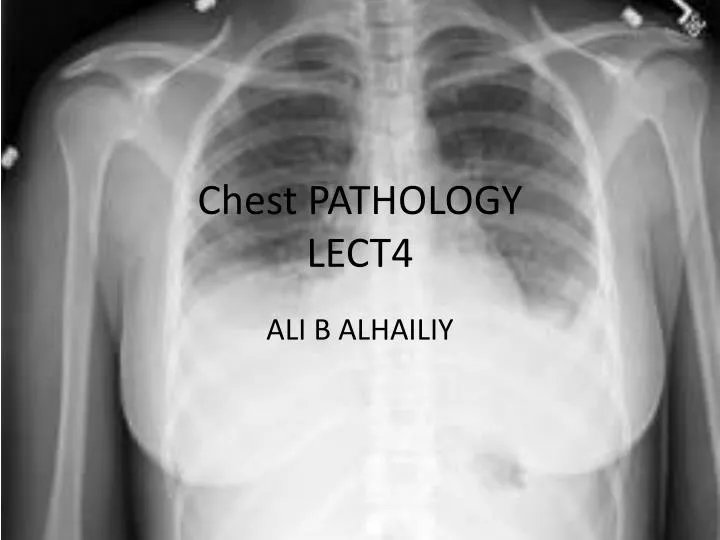
Chest PATHOLOGYLECT4 ALI B ALHAILIY
1- Pneumonia • Pneumonia is an inflammatory condition of the lung—affecting primarily the microscopic air sacs known as alveoli. It is usually caused by infection with viruses or bacteria and less commonly other microorganisms .
Signs and symptoms • a productive cough • fever accompanied by shaking chills • shortness of breath • sharp or stabbing chest pain during deep breaths • increased respiratory • The typical signs and symptoms in children under five are fever, cough, and fast or difficult breathing.
DIAGNOSIS • Pneumonia is typically diagnosed based on a combination of 1- physical signs and 2- a chest X-ray. However, the underlying cause can be difficult to confirm, as there is no definitive test able to distinguish between bacterial and non-bacterial origin.
A- Physical examination may sometimes reveal low blood pressure, high heart rate, or low oxygen saturation. The respiratory rate may be faster than normal, and this may occur a day or two before other signs • B- Imaging • A chest radiograph is frequently used in diagnosis. • In people with mild disease, imaging is needed only in those with potential complications, those not having improved with treatment, or those in which the cause is uncertain
2- Pulmonary embolism (PE) • Pulmonary embolism (PE) is a blockage of the main artery of the lung or one of its branches by a substance that has travelled from elsewhere in the body through the bloodstream (embolism). PE most commonly results from deep vein thrombosis (a blood clot in the deep veins of the legs or pelvis) that breaks off and migrates to the lung
Sign and Symptoms • Symptoms of pulmonary embolism include • difficulty breathing • chest pain on inspiration • Clinical signs include low blood oxygen saturation • Cyanosis • rapid breathing • rapid heart rate. • Severe cases of PE can lead to collapse, abnormally low blood pressure, and sudden death.
Diagnosis • The gold standard for diagnosing pulmonary embolism (PE) is CT scans . • Pulmonary angiography is used less often due to wider acceptance of CT scans, which are non-invasive. • CT pulmonary angiography is the recommended first line diagnostic imaging test in most people.
Acquisition • An intravenous cannulais required for the administration of the 50-150 ml of Contrast media. • This is injected, usually automatically, by a power injectorat a rate of up to 5 ml/s. • Many hospitals use bolus tracking, where the scan commences when the contrast is detected at the level of the proximal pulmonary arteries. • If this is done manually, scanning commences about 10–12 seconds after the injection has started. • Slice thickness will be around 1.25mm up to 2.5mm • Modern CT scanners with a scan rate of up to 320 mm/s can acquire all the images within a 1 second X-ray exposure, avoiding the problems of respiratory motion, cardiac motion and contrast draining from the pulmonary circulation during the study.
Interpretation • On CTPA, the pulmonary vessels are filled with contrast, and appear white. • Any mass filling defects (embolus or other matter such as fat) appears darker. • Ideally, the scan should be complete before the contrast reaches the left side of the heart and the aorta, as this may mean contrast has drained from the pulmonary arteries, or require a larger dose of contrast media.
PE With contrast (CTPA) Without contrast
A large pulmonary embolism at the bifurcation of the pulmonary artery
CT pulmonary angiography (CTPA) showing a "saddle embolus" at the bifurcation of the pulmonary artery
3-Pulmonary consolidation • A pulmonary consolidation is a condition whereby the lung tissues solidify because of the accumulation of solid and liquid material in the air spaces. • The major cause of this is pneumonia. • Other causes include a collapsed lung, tumours of the lung and infections such as ascariasis, syphilis . • The fluid can be pulmonaryedema, pus, inhaled water, or blood (from bronchial tree or hemorrhage from a pulmonary artery. • It is clinically important in pneumonia: the signs of lobar pneumonia are characteristic and clinically referred to as consolidation
Diagnosis by Radiology • Typically, an area of white lung is seen on a standard X-ray. Consolidated tissue is more radio-opaque than normally aerated lung parenchyma, so that it is clearly demonstrable in radiography and on CT scans. • Consolidation is often a middle-to-late stage feature/complication in pulmonary infections.
Pneumonia as seen on chest X-ray. A: Normal chest X-ray. B: Abnormal chest X-ray with consolidation from pneumonia in the right lung, middle or inferior lobe (white area, left side of image).
Lung cancer • Lung cancer is cancer that starts in the lungs. • There are two main types of lung cancer: • Non-small cell lung cancer (NSCLC) is the most common type of lung cancer. • Small cell lung cancer (SCLC) makes up about 20% of all lung cancer cases. • If the lung cancer is made up of both types, it is called mixed small cell/large cell cancer. • If the cancer started somewhere else in the body and spreads to the lungs, it is called metastatic cancer to the lung.
causes • Lung cancer is the deadliest type of cancer for both men and women. • Lung cancer is more common in older adults. It is rare in people under age 45. • Cigarette smoking is the leading cause of lung cancer. • Lung cancer can also affect persons who have never smoked. • Secondhand smoke (breathing the smoke of others) increases your risk of lung cancer. • The following may also increase your risk of lung cancer: • Exposure to asbestos • Exposure to cancer-causing chemicals such as uranium, beryllium, vinyl chloride, nickel chromates, coal products, mustard gas, chloromethyl ethers, gasoline, and diesel exhaust • Exposure to radon gas • Family history of lung cancer • High levels of air pollution • High levels of arsenic in drinking water • Radiation therapy to the lungs
Symptoms • Early lung cancer may not cause any symptoms. • Symptoms depend on the type of cancer you have, but may include: • Chest pain • Cough that does not go away • Coughing up blood • Fatigue • Losing weight without trying • Loss of appetite • Shortness of breath • Wheezing
Exams and Tests • 1- physical exam and history • 2-chest x ray • 3- complete blood count (CBC) • 4- CT Scan • 5- Positron emission tomography (PET) scan
Chest CT Scan Lung window Soft tissue window