Download
1 / 38
480 likes | 1.39k Views
Miscarriage Early pregnancy loss. Dr Chro Najmaddin Fattah MBChB, DGO, MRCOG, MRCPI, MD Obst. & Gyne. Department. Definition:. Spontaneous miscarriage is the most common complication of early pregnancy before 24 week gestation 8–20% clinically recognized pregnancies 13–26% all pregnancies

E N D
Miscarriage Early pregnancy loss Dr Chro Najmaddin Fattah MBChB, DGO, MRCOG, MRCPI, MD Obst. & Gyne. Department
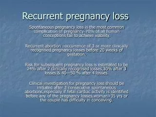
Definition: Spontaneous miscarriage is the most common complication of early pregnancy before 24 week gestation8–20% clinically recognized pregnancies13–26% all pregnancies Incidence: 15% Early pregnancy loss: If it occurs before 12 weeks (80%) Late pregnancy loss: If it occurs between 13 to 24 weeks (12%) ( usually there is a fetus)
Early pregnancy loss classified into; No fetus on U/S examination (Empty gestational sac) Fetal tissues absent on histological examination Early fetal demise: fetus present on U/S examination fetal tissues present on histological examination Factors influence rate of spontaneous miscarriage: • Maternal age > 35 years • Gravidity • Previous miscarriage • Multiple pregnancies
Ultrasound Findings of EPL Anembryonic Pregnancy — No fetal pole with mean sac diamter >25 mm (transabdominal) OR >18 mm (transvaginal) — <4 mm growth in 7 days(No yolk sac, with mean sac diameter >25mm) Embryonic Demise — No cardiac activity with CRL ≥7mm Mishell DR, Comprehensive Gynecology 2007
Etiology: • Abnormal conceptus as genetic abnormalities (50-60%), • structural abnormalities • Endocrine abnormalities (10- 15%) • Cervical incompetence (8-10%) • Uterine anatomic abnormalities (1-3%) • Immunological (5%) • Infections (3-5%) • Structural abnormalities • Unknown reasons (< 5%)
Abnormal conceptus Means an empty gestational sac without embryo development. (Blighted ovum ) Most miscarriage occurs before 8 weeks’ gestations. Result from: Error in maternal and/ or paternal meiosis chromosomal division without cytoplasmic division
The abnormalities of development may be due to: Chromosomal abnormalities Structural abnormalities Gene defects (absence of specific enzyme) I-The chromosomal abnormalities; Are found in approximately 80% of empty sac( blighted ovum) and 5-10% of the miscarriage in which the a fetus is present. These are the most frequent and important causes of early pregnancy loss
The chromosomal abnormalities include; ♣ Autosomal trisomy; The non-disjunction defect is found approximately in 60% of blighted ovum with abnormal karyotypes. most non-disjunction occurs during 1st mitotic division The affected chromosomes are: 16 (32%) 22 (10%) 21 (8%) ♣ Triploidy ; occurs in 12-15% of chromosomal abnormalities double paternal chromosomes (69 chromosomes) partial molar of pregnancy occurs in 5% ♣ Monosomy X; represents 25% of miscarriage with chromosomal abnormalities (45X)
♣Structural rearrangement; the abnormality consists of unbalanced translocation accounts 3-5% of miscarriage with abnormal chromosome 3% of couple s will be carrier karyotyping is required II- structural abnormalities as Nural tube deffect (NTD) , uncommon cause of miscarriage III- Gene defect; -difficult to determine because of facilities to identify the individual gene defects. -Example as autosomal dominant disorders and X-linked dominant disorders.
II- Endocrine causes *Corpus luteum is essential for maintenance of pregnancy during the first 8 weeks. * Surgical removal of it→ miscarriage within 4- 7 days * Parenteral progesterone may prevent miscarriage but the evidence of progesterone deficiency as a cause of miscarriage is unsatisfactory. * In the past, progesterone have been used among women with recurrent miscarriage with good results. It is possible that corpus luteum deficiency could be a cause of early pregnancy loss * Use pf progesterone is over used in miscarriage.
III-Uterine abnormalities A- Uterine malformations; Result from a failure of normal fusion of the Mullerian ducts, as: bicronuate uterus, septate or subseptate, and uterus didelphys. May result in miscarriage in 10- 15% B- Intra-uterine synechiae ( Asher man's syndrome) in which there is either partial or complete adhesion between walls of uterus leading to partial or complete obliteration of the uterine cavity. Usually occur as a result of intrauterine infections following; Retained parts of conception post-abortal or postpartum curettage Repeated pregnancy loss
C- Cervical incompetence Is a well recognized cause of miscarriage in late second trimester ▲ The clinical feature are: - painless cervical dilatation (main presentation) - increase vaginal discharge - speculum examination shows bulging membrane with cervical dilatation ▲Causes; Trauma to cervix is the main etiological factor - vigorous mechanical dilatation of cervix - trauma during delivery - cone biopsy - cervical amputation Congenital; rare
▲ Diagnosis of cervical incompetence 1- History and examination 2- During pregnancy: U/s examination Finding: short cervix internal os dilated up to ≥ 2cm funnel shaped cervix 3- Non pregnancy: passing Hegar dilator number 8 through internal os hysterosalpingography
▲ Treatment Placing suture ( cervical cerclage) around the cervix at 14- 16 week’s gestation Two types of sutures; McDonald Shrodkar ▲ Complications of cerclage - Rupture of membrane - Infections - further trauma to cervix ▲ Time of removal of cerclage at 38 weeks
D- Infection ◙ uncommon cause of miscarriage ◙ acute maternal infections as ; peyelitis, appendicitis can lead to general toxic illness with high temperature that stimulates the uterine activity → miscarriage. ◙ early diagnosis & treatment will control most of infection and forestall the occurrence of miscarriage ◙ syphilis can cross the placenta → IUFD and miscarriage ◙ other infections as; Rubella, Toxoplasmosis, Listeriosis, CMV, and Mycoplasma can lead to miscarriage
E-Immunological causes • Immunological rejection of fetus can cause recurrent miscarriage • May be due to failure of the normal immune response in mother • An example is anti-phospholipids antibody syndrome responsible for 3-5% of recurrent miscarriage F- toxic factors Anesthetic gases, smoking, alcohol, and drug abuse can cause miscarriage G- Trauma amniocentesis, CVS, IUCDs, and abdominal surgery
Types of miscarriage 1- Threatened miscarriage Referred as vaginal bleeding before 24 week’s gestation when there is a viable fetus without evidence of cervical dilatation and pain. 2- Inevitable, if the cervix becomes dilated, the bleeding increases and there is pain. 3- Incomplete, if there is partial expulsion of product of product of conception (usually the fetus) with retention of some parts ( usually placenta). 4- Complete, complete expulsion of product of conception. 5- Missed miscarriage, the embryo dies in utero but is not passed 6 -Septic, infection may occur following any type of abortion and may spread to pelvis or even leads to septicemia.
7- Recurrent miscarriage, referred as three or more consecutive miscarriage Clinical features of miscarriage 1- Threatened miscarriage - vaginal bleeding (usually slight) - slight abdominal cramps - internal os is closed - viable fetus on U/S examination 2- Inevitable miscarriage - bleeding becomes heavy with clots - lower abdominal pain - cervix dilated ± bulging membrane
3- Incomplete miscarriage - heavy vaginal bleeding may lead to hypo-volaemic shock - lower abdominal pain some times sever - history of passing something (POC) - cervix dilated - Retained parts of conception on U/S examination 4- Complete miscarriage - bleeding minimal - no pain - cervix closed - empty uterus on U/S examination
Differential diagnosis • Ectopic pregnancy • Hydatiform mole ( molar pregnancy) • Local causes as; cervical erosion, cervical polyp, etc. Clinical assessment A- History; includes personal history complains as; vaginal bleeding, pain medical history
B- Examination * General assessment for any signs of shock * Abdominal examination for: abdominal tenderness size of uterus large: wrong date multiple pregnancy molar pregnancy fibroids smaller : wrong date non- viable fetus
* Pelvic examination Should be carried out in all cases If the vaginal bleeding is slight → speculum examination for - any vaginal infection - cervical lesion If the bleeding is heavy → digital examination to assess - cervical tenderness ? Ectopic - state of cervix - any RPOC felt inside cervix ↓ to be removed manually ↓ relieve pain & decrease bleeding
C- Investigation • Serum B-HCG may be required to confirm pregnancy • Ultra-sound examination Abdominal U/S GS will be seen normally if SBHCG ≥ 3000mIU/ml Trans-vaginal ; more accurate GS will be seen normally if SBHCG ≥ 1500mIU/ml NB; if fetal heart seen on U/S examination, pregnancy will continue in 98%.
Management Options Do Nothing: Expectant management Do Something: Medical management Do Surgery: Surgical management Sotiriadis A, Obstet Gynecol 2005Nanda K, Cochrane Database Syst Rev 2006
Comparison of Outcome by MethodManagement of Early Pregnancy Loss FactorComparison of Methods Success rate Surgical > Medical Medical ≥ Expectant Resolution Surgical > Medical > Expectant within 48 hrs Infection risk Expectant = Medical = Surgical.2–3% Nanda K, Cochrane Database Syst Rev 2006; Nielsen S, Br J Obstet Gynaecol 1999; Shelly JM, Aust. NZ J Obstet Gynaecol 2005; Sotiriadis A, Obstet Gynecol 2005; Tinder J, (MIST) BMJ, 2006
Do Nothing: Expectant Management Overall success rate 81% Success rates vary by type of miscarriage— Incomplete/inevitable abortion 91%— Embryonic demise 76%— Anembryonic pregnancies 66% Luise C, Ultrasound Obstet Gynecol 2002
Medical Management Success Rates Placebo 16–60% Single dose misoprostol 25–88% 400–800 mcg Repeat dose x 1 if incomplete 80–88% at 24 hours • Success rate depends on type of miscarriage — 100% with incomplete abortion — 87% for all others Wood SL, Obstet Gynecol 2002; Bagratee JS, Hum Reproduct 2004; Blohm F, BJOG: Int J Obstet Gynecol 2005
Medical ManagementRequirement for Therapy <13 weeks gestation Stable vital signs No evidence of infection No allergies to medications used Adequate counseling and patient acceptance of side effects
Misoprostol Prostoglandin E1 analogue FDA approved for prevention of gastric ulcers Used off-label for many Ob/Gyn indications:— Labor induction— Cervical ripening— Medical Miscarriage (with mifepristone)— Prevention/treatment of postpartum hemorrhage Can be administered by oral, buccal, sublingual, vaginal and rectal routes Chen B, Clin Obstet Gynecol 2007
Surgical ManagementEarly Pregnancy Loss Suction dilation and curettage (D&C) Who should have surgical management?— Unstable— Significant medical morbidity— Infected— Very heavy bleeding— Anyone who WANTS immediate therapy
Management 1- Threatened miscarriage - Reassurance of patients - Rest for few days until the bleeding has settled down - May require progesterone supplementation - Folic acid 2- Incomplete miscarriage - Assessment of general condition - Blood sample for blood group, RH factor, and CBC - Removal of RPOC if felt in cervical canal - Ergometrine 0.5mg IV or IM to ↓ blood loss
- Evacuation of uterus UGA followed by gentle curettage - Ergometrine 0.5mg IV will encourage uterine contraction -Anti D if RH negative - If there is hypo-volaemic shock, may require blood transfusion Septic miscarriage Occurs as a result of ascending infection following miscarriage. If not treated, infection may spread throughout pelvis → septicemia and septic shock Signs; pyrexia abdominal pain, and tenderness persistent vaginal bleeding offensive vaginal discharge
Investigation • Routine basic investigations as BL. Group, RH factor, CBC, BS, urea & electrolytes, etc • Cervical swab • U/S examination for retained parts Treatment • Iv. Broad spectrum antibiotic • IV fluids ± blood transfusion if needed • Analgesia • Evacuation of uterus • Anti D
Complications of septic miscarriage • Septicemia, and septic shock • Acute renal failure • Chronic pelvic infection • Infertility Missed miscarriage clinical feature: - Disappearance of symptoms of pregnancy -Size of uterus < duration of gestation - U/S shows no signs of fetal life -PT will remains positive as long as the placental tissues survive then → -ve Treatment: there is no urgency in treating missed miscarriage because: spontaneous miscarriage mostly occurs coagulation defects due to dead fetus syndrome are rare
Recurrent miscarriage Management includes: 1-Careful history and examination 2- trans-vaginal U/S 3- HSG and/or hysteroscopy 4- karyotyping 5-blood tests for infections 6- antiphospholipid antibodies Treatment according to the cause
Induced abortion Induced abortion is not considered in medical terms alone but it arouses strong personal emotions and involves religious and ethical considerations. Indications; termination of pregnancy may be medically indicated to safe life of patients as in: malignant diseases of cervix, breast and sever cardiac disease. Also fetal malformation may require termination.
Q question : 1- what is miscarriage and the types? 2- how to diagnose different types of miscarriage ? 3 what are the complications ? How to treat patient ?


![[PDF] The Miscarriage Map Workbook: An Honest Guide to Navigating Pregnancy Loss](https://cdn7.slideserve.com/12515934/slide1-dt.jpg)