Download
1 / 56
560 likes | 572 Views
Lymphatic System. Vessels, Organs and Tissues. Lymphatic System. Vessels: Connect to CV Lymphatics Capillaries Collecting vessels Trunks Transport fluid => lymph Organs: house immune cells LN Spleen Thymus Tissues Tonsils GALT MALT. Fluid Compartments. Capillary Dynamics

E N D
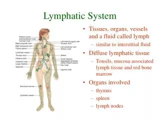
Lymphatic System Vessels, Organs and Tissues
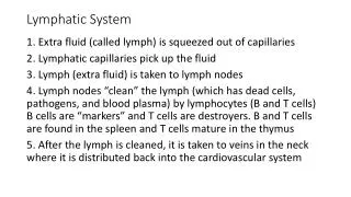
Lymphatic System • Vessels: Connect to CV • Lymphatics • Capillaries • Collecting vessels • Trunks • Transport fluid => lymph • Organs: house immune cells • LN • Spleen • Thymus • Tissues • Tonsils • GALT • MALT
Fluid Compartments • Capillary Dynamics • Hydrostatic/Hydraulic • Osmotic • Fluid Compartments • ICF • ECF • ISCF • Plasma • Lymph
Lymph Node: Gross Anatomy • 1. Afferent Lymphatics • 2. Cortex • 3. Germinal Center • 4. Capsule • 5. Medullary cord • 6. Valve • 7. Efferent Lymphatics
LN Structure: CT and vessels • Capsule • Subcapsular sinus • Trabecula • Reticular CT stroma • Vessels • Types • Afferent • Efferent • Valves
Lymph Node: Histology B cells T cells MO
Lymph Node: Cortex Lymphatic Nodule Germinal Center
Lymph Node: Medullary Cords Medullary Cords Sinusoids Macrophages
L.N. Medullary Cells • P = Plasma cells (daughter cells of Activated B cells • M = Macrophages (mature monocytes in tissues)
Diagnostics for Lymphatics and LN Lymphangiogram
Spleen: Histology Splenic Sinusoid
Spleen Problems • Rupture • Cancer • Infection Hydatid cyst
Thymus Embryological Development • 1. Mandibular arch • 2. Hyoid arch • 3. Cervical sinus entry • 4. 3rd pharyngeal pouch • 5. 4th pharyngeal pouch • 6. Foramen cecum • 7. Thyroid • 8. Cervical sinus • 9. Thymus (3rd pouch) • 10. Thymus (4th pouch)
Thymic Cell Development • 1. Thymic capsule • 2. Thymic nurse cells • 3. CT septa and BV • 4. Subcapsular epithelium (for the blood thymus barrier) • 5. Cortical Epithelial cells • 6. Medullary epithelial cells • 7. Dendritic cells • 8. Hassall’s Corpuscle • 9. Macrophages • 10. Cortex • 11. Medulla
Thymic Problem Thymic Aplasia
Tonsil: Location • Pharyngeal [Adenoids] • Palantine • Lingual • Tubal
Ileum Problems Inflammation Necrosis