Download
1 / 1
10 likes | 114 Views
E13 Hu ChAT. P12. P30. P0. P3. E15 Hu ChAT. Spatio-temporal differentiation of cholinergic neurons during enteric nervous system development. Scott Lee 1 , Chris Erickson 1 , Amanda Barlow 2 , and Miles L. Epstein 1 .

E N D
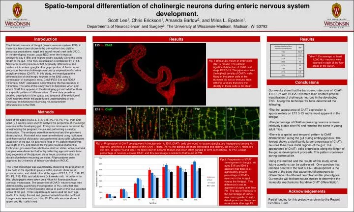
E13 Hu ChAT P12 P30 P0 P3 E15 Hu ChAT • Spatio-temporal differentiation of cholinergic neurons during enteric nervous system development. • Scott Lee1, Chris Erickson1, Amanda Barlow2, and Miles L. Epstein1. Departments of Neuroscience1 and Surgery2, The University of Wisconsin-Madison, Madison, WI 53792 Introduction Results Results The intrinsic neurons of the gut (enteric nervous system, ENS) in mammals have been shown to be derived from two distinct precursor populations: vagal and sacral neural crest cells (NCC). In the developing mouse, vagal NCC enter the foregut at embryonic day 9 (E9) and migrate rostro-caudally along the entire length of the gut. This NCC colonization is completed by E14.5. NCC form neural precursors that eventually differentiate and coalesce into enteric ganglia. A large proportion of these neural precursors become cholinergic neurons by expression of choline acetyltransferase (ChAT). In this study, we investigated the differentiation of cholinergic neurons in the ENS using a combination of transgenic mice, ChAT-IRES-Cre and ROSA TdTomato. ChAT expression is identified by the fluorescence of TdTomato. The aims of this study were to determine when and where ChAT first appears in the developing gut and whether there is a specific pattern of differentiation. These data provide a detailed description of the spatial and temporal differentiation of ChAT neurons which will guide future understanding of the molecular mechanisms influencing neurotransmitter differentiation in the ENS. Table 1: On average, at least 1,000 Hu+ neurons were counted in each of the four areas of the gut on. Fig. 1 Whole-gut mount of embryonic-day 13 mouse. The earliest significant detection of ChAT is at stage E12.5-13. The jejunum shows the highest density of ChAT+ cells. Many of the green cells in the stomach did not co-immuno stain with Hu, and are therefore the identity of these cells is not clear. Conclusions • Our results show that the transgenic intercross of ChAT-IRES-Cre with ROSA TdTomato mice enables precise visualization of cholinergic neurons in the developing ENS. Using this technique we have determined the following: • The first appearance of ChAT expression is approximately on E12.5-13 and is most apparent in the foregut. • The percentage of ChAT-expressing neurons remains relatively stable after P6 and remains the same in young adult mice. • There is a spatial and temporal pattern to ChAT differentiation along the gut during embryogenesis. The foregut shows a significantly larger percentage of ChAT+ neurons than more distal regions of the gut. The appearance of ChAT+ cells progresses along the length of the gut as development proceeds. This pattern continues during postnatal life. • Using this method and the results of this study, other future questions can be addressed. One question that remains central to the field of ENS development is the nature of the cues that cause neural precursors to differentiate into different neurotransmitter phenotypes. Our results will facilitate future studies to determine the molecular mechanisms that drive ChAT differentiation. Methods Mice at the ages of E12.5, E15, E16, P0, P3, P6, P12, P30, and adult (< 6 weeks) were used to analyze the proportion of cholinergic neurons in the developing gut. Embryonic mice were harvested by anesthetizing the pregnant mouse and performing a cervical dislocation. The embryos were then removed and the guts were dissected out. Postnatal mice were anesthetized using isoflurane and then cervical dislocation was performed along with subsequent dissection of the gut. All guts were fixed in 4% paraformaldehyde overnight at 4oC and stained for the pan neuronal marker Hu. Embryonic guts were then whole-mounted on slides, while postnatal samples were dissected further by collecting approximately 1cm-long segments of the jejunum, distal ileum, proximal colon, and distal colon before mounting on slides. All procedures were approved by University of Wisconsin-Madison IACUC. The ChAT phenotype was quantified by observing the proportion of Hu+ cells in the myenteric plexus in the jejunum, distal ileum, proximal colon, and distal colon at the ages of E12.5, E15, E16, P0, P3, P6, P12, P30, and adult mice (< 6 weeks old). In order to do this, photographs were taken on a Nikon A1 fluorescent laser confocal microscope. The proportion of ChAT+ neurons was then determined by quantifying the proportion of Hu+ cells that also expressed ChAT in the myenteric plexus of each of the four selected areas of the gut. Three separate guts were used for each age (n=3). For clarity, the red and green channels from the confocal images were reversed, such that ChAT+ cells are now shown in green and Hu+ cells in red. . Fig. 2: Progression of ChAT development in the jejunum. At E15, ChAT+ cells are found in nascent ganglia, are interspersed among Hu+ neurons, and there is a presence of thin ChAT+ fibers. At P0, the ganglia are more developed and distinct, but the ChAT+ fibers are still thin. At ages P3 and older, the fibers start to become thicker and reach other ganglia to form connections. At P12, a large percentage of neurons express ChAT, and this percentage is similar to that found at P30. Fig 3: Progression of ChAT development in the gut. At ages E15 and E16 it appears that there is a significantly greater percentage of ChAT+ neurons in the foregut versus the hindgut. This difference is not as apparent at ages later than P0. The rate of increase in the percentage of ChAT+ neurons is greater in early development and becomes more stable after age P6. Acknowledgements Partial funding for this project was given by the Regent Scholars Fund.