Download
1 / 40
470 likes | 613 Views
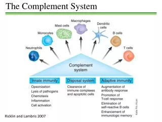
The complement system. Complement functions in two (three?) systems: Alternative Classical Lectin-based. Complement has three functions: Opsonin Chemoattractant Membrane Attack Complex (MAC). The complement system.

E N D
Complement functions in two (three?) systems: Alternative Classical Lectin-based • Complement has three functions: • Opsonin • Chemoattractant • Membrane Attack Complex (MAC)
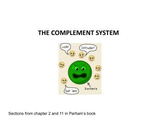
The complement system • A defensive system consisting of over 30 proteins produced by the liver and found in circulating blood serum. • Complement kills microbes in three different ways • 1. opsonization • 2. inflammation • 3. Cytolysis
Complement refers to a complex set of 14 distinct serum proteins (nine components) that are involved in three separate pathways of activation. • Components numbered in order of discovery. • Sequence of activation is not in numerical order. • Components circulate in inactive precursor form, develop activity upon activation. • Complement proteins designated by “C” followed by numbers and letters.
Promote the inflammatory response by opsonization which enhances susceptibility of coated cells to phagocytosis. • Alter biological membranes to cause direct cell lysis
General Properties of Complement • Primary role is cell lysis. • Activity of complement destroyed by heating sera to 56 C for 30 minutes. • IgM and IgG are the only immunoglobulin capable of activating complement (classical pathway). • Complement activation can be initiated by complex polysaccharides or enzymes (alternative or properdin pathway). • Portions of the complement system contribute to chemotaxis, opsonization, immune adherence, anaphylatoxin formation, virus neutralization, and other physiologic functions
A Cascade system • The complement works as a cascade system. • Cascade is when one reaction triggers another reaction which trigger others and so on. These types of systems can grow exponentially very fast.
Cascade activation • Complement proteins are often designated by an uppercase letter C and are inactive until they are split into products. • Example: C1 • When the products are split they become active. The active products are usually designated with a lower case a or b. • Example: C1a and C1b
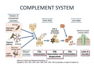
Two Pathways • The complement pathway can be activated by either of two different pathways. • Classical pathway (specific immune system) • alternative (non-specific immune system)
The Classical Pathway • The classical pathway is considered to be part of the specific immune response because it relies on antibodies to initiate it. • C1 becomes activated when it binds to the ends of antibodies
C1: The Recognition Unit • C1 consists of 3 subunits: C1q, C1r, and C1s. • C1q molecule consists of a collagenous region with six globular head groups globe end serves as recognition unit • When antibody binds to antigen, binding sites for the globular head groups of C1q are exposed on the Fc region of the antibody. • For C1q to initiate the cascade it must attach to at least 2 Fc fragments, requires at least 2 molecules of IgG or one molecule of IgM. • C1q binding causes C1r to enzymatically activate C1s.
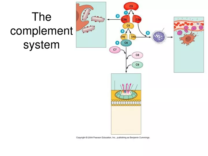
The Activation Unit (C4b2a3b) C1s cleaves C4 into C4a and C4b C1s cleaves C2 into C2a and C2b C4b2a (C4b2b in some texts) is enzymatically active and can cleave many molecules of C3 into C3a and C3b.
Membrane Attack Unit (C5,6,7,8,9) In the presence of C5b, molecules of C6, C7, C8 and a variable number of C9 molecules assemble themselves into aggregates. This molecular complex causes a change in membrane permeability. Exact cause of lysis unknown, one theory is change in lipid membrane causes exchange of ions and water molecules across membrane. Cells can lyse without C9 but it’s slower.
The building of a C3 activation complex • Once C1 is activated, it activates 2 other complement proteins, C2 and C4 by cutting them in half • C2 is cleaved into C2a and C2b • C4 is cleaved into C4a and C4b • Both C2b and C4b bind together on the surface of the bacteria • C2a and C4a diffuse away
C3 Activation complex • C2b and C4b bind together on the surface to form a C3 activation complex • The function of the C3 activation complex is to activate C3 proteins. • This is done by cleaving C3 into C3a and C3b
C3b • Many C3b molecules are produced by the C3 activation complex. • The C3b bind to and coat the surface of the bacteria. • C3b is an opsonin • Opsonins are molecules that bind both to bacteria and phagocytes • Opsonization increases phagocytosis by 1,000 fold. Bacteria Opsonins
C3a C3a increases the inflammatory response by binding to mast cells and causing them to release histamine
Building the C5 activation complex • Eventially enough C3b is cleaved that the surface of the bacteria begins to become saturated with it. • C2b and C4b which make up the C3 activation complex has a slight affinity for C3b and C3b binds to them • When C3b binds to C2b and C4b it forms a new complex referred to as the C5 activation complex
The C5 activation complex • The C5 activation complex (C2b, C4b, C3b) activates C5 proteins by cleaving them into C5a and C5b • Many C5b proteins are produced by the C5activation complex. These C5b begin to coat the surface of the bacteria.
The function of C5a • C5a disperses away from the bacteria. • Binds to mast cells and increases inflammation. • Most powerful chemotactic factor known for leukocytes
Building the Membrane Attack complex • C5b on the surface of bacteria binds to C6 • The binding of C6 to C5b activates C6 so that it can bind to C7 • C7 binds to C8 which in turn binds to many C9’s • Together these proteins form a circular complex called the Membrane attack complex (MAC)
Membrane Attack complex • The MAC causes Cytolysis. • The circular membrane attack complex acts as a channel in which cytoplasm can rush out of and water rushes in. • The cells inner integrity is compromised and it dies • Animation of the classical pathaway
The alternative pathway • The alternative pathway is part of the non-specific defense because it does not need antibodies to initiate the pathway. • The alternative pathway is slower than the Classical pathway
Alternative Pathway (Properdin Pathway) • Cleavage of C3 and activation of the remainder of the complement cascade occurs independently of antibody. • Triggers for the alternative pathway include • Bacterial cell walls • Bacterial lipopolysaccharide • Fungal cell walls • Virally infected cells • Rabbit erythrocytes
Alternative Pathway (Properdin Pathway) Molecules of C3 undergo cleavage at continuous low level in normal plasma. At least 4 serum proteins (factor B, factor D, properdin (P), and initiating factor (IF) function in this pathway. C3b attaches to appropriate site (activating surface) which is actually a protective surface
Alternative Pathway (Properdin Pathway) Action by the 4 serum proteins on C3b proceeds to the C3 activator stage without participation of C1, C4 or C2. Activation sequence: C3, C5, C6, C7, C8, C9.
Initiation of The Alternative pathway • C3 contains in unstable thioester bond. • This unstable bond makesC3 subject to slow spontaneous hydrolysis to C3b and C3a • The C3b is able to bind to foreign surface antigens. • Mammalian cells which inactivates C3b
Factor B • C3b on the surface of a foreign cells binds to another plasma protein called factor B
Factor D • The binding of C3b to factor B allows a protein enzyme called Factor D to cleave Factor B to Ba and Bb. • Factor Bb remains bound to C3b while Ba and Factor D disperse away.
The C3 activation complex • Properdin, also called factor P, binds to the C3bBb complex to stabilize it. • C3bBbP make up the C3 activation complex for the alternative pathway
The C3 activation Complex • The C3 activation complex causes the production of more C3b. • This allows the initial steps of this pathway to be repeated and amplified • 2X10⁶ molecules can be generated in 5 minutes
C5 activation complex • When an additional C3b binds to the C3 activation complex it converts it into a C5 activation complex. • The C5 activation complex cleaves C5 into C5a and C5b. • C5b begins the production of the MAC.
Lectin Pathway Activation of the lectin pathway begins when mannan-binding protein (MBP) binds to the mannose groups of microbial carbohydrates. Two more lectin pathway proteins called MASP1 and MASP2 (equivalent to C1r and C1s of the classical pathway) bind to the MBP. This forms an enzyme similar to C1 of the classical complement pathway that is able to cleave C4 and C2 to form C44bC2a, the C3 convertase capable of enzymatically splitting hundreds of molecules of C3 into C3a and C3b
Lectin Pathway • The beneficial results are the same as in the classical complement pathway: • Trigger inflammation (C5a>C3a>C4a); • Chemotactically attract phagocytes to the infection site (C5a); • Promote the attachment of antigens to phagocytes via enhanced attachment or opsonization (C3b>C4b; • Serves as a second signal for the activation of naive B-lymphocytes (C3d); • Cause lysis of gram-negative bacteria and human cells displaying foreign epitopes (MAC). • And remove harmful immune complexes from the body (C3b>C4b).
C3 convertase C3b C3 C3b Activation of the Lectin Pathway mannan binding lectin (MBL) mannose sugars MBL covalent bond with surface “activating surface” (bacterial or yeast cell surface) -mannan binding lectin (MBL) recognizes mannose sugars on microbial cells -host mannose is hidden and is not accessible to MBL -lectin pathway results in the formation of a C3 convertase that generates C3b -C3b forms a covalent bond with the surface of the pathogen and is part of C5 convertase