Download

1 / 67
910 likes | 3.78k Views
Approach to Mediastinal Masses . Clinical Presentation: 33% of all masses present in patients less than 15 years old If small, usually asymptomatic and found incidentally (cautious work up) If large, usually present with respiratory distress (frantic work up) .

E N D
Clinical Presentation:33% of all masses present in patients less than 15 years oldIf small, usually asymptomatic and found incidentally (cautious work up)If large, usually present with respiratory distress (frantic work up)
-The anterior mediastinal compartment is bordered by the sternum anteriorly, and the ventral cardiac surface posteriorly. -This compartment contains fat, ascending aorta, lymph nodes, internal mammary artery and vein, adjacent osseous structures (ribs and sternum), thymus. -Therefore will most likely see masses typical to these structures, ie a lymphoma in lymph nodes.
It is located above a horizontal line drawn from the angle of Louis posteriorly to the spine. Structures in the superior mediastinal compartment include the thyroid gland, aortic arch and great vessels, proximal portions of the vagus and recurrent laryngeal nerves, esophagus and trachea.
The borders are composed of the anterior mediastinal compartment ventrally, and the anterior surface of the spine, posteriorly. Structures in the middle mediastinal compartment include the esophagus (which will not be visible unless there is a problem), vagus nerve, recurrent laryngeal nerve, heart, proximal pulmonary arteries and veins (hilar), trachea and root of the bronchial tree, and superior and inferior vena cava
The posterior mediastinum borders the anterior surface of the spine posteriorly to the ribs. Structures in the posterior mediastinal compartment include the descending aorta, adjacent osseous structures (the spine and ribs) and nerves, roots, spinal cord, and the azygous and hemiazygous veins.
Anterior Mediastinal Masses: (4 T's) (30% of mediastinal masses) • Thymoma • Teratoma • Thyroid (Ectopic) • (Terrible) Lymphoma
Middle Mediastinal Masses (A + B) (30% of mediastinal masses) • Adenopathy (infection [bacterial, granulomatous], neoplasm [leukemia / lymphoma, metastases]) • Bronchopulmonary foregut malformations (Esophageal duplication cyst, bronchogenic cyst, sequestration)
Posterior Mediastinal Masses: (N) (40% of mediastinal masses) • Sympathetic ganglion tumors: neuroblastoma, ganglioneuroblastoma, ganglioneuroma (95% of posterior mediastinal masses) • Neurofibroma • Neurenteric cyst • Extramedullary hematopoesis • Paravertebral soft tissue mass from infection
Approach/Discussion: • PA and lateral chest films are the first step in distinguishing from which mediastinal compartment the mass is arising from. • Computed tomography or magnetic resonance imaging is the next step, better characterizing the nature and extent of the lesion, thus narrowing the differential diagnosis. MRI is especially good at looking for spinal canal invasion in posterior mediastinal masses • Tissue biopsy is required for definitive diagnosis, and surgical resection for definitive cure.
PA and lateral chest films show a large anterior mediastinal mass causing narrowing and rightward deviation of the trachea. The mass is not calcified.
CT exam show a low density mass in the anterior mediastinum with irregular walls with calcium in it. Dx Teratoma, Anterior Mediastinal
single slice from an enhanced chest CT exam shows the mass to be non-enhancing, posterior to the right bronchi, and next to the esophagus. Dx: Esophageal Duplication
Eighteen year old female with an incidentally noted chest mass
Eleven year old male with upper respiratory symptoms and wheezing.
Slice from an enhanced chest CT exam shows a multi-loculated non enhancing mass in the anterior mediastinum Dx-Thymic Cyst
Soft tissue in the anterior mediastinum compatible in appearance with thymus
Twelve year old female with a chest mass PA and lateral chest films show a large, lobulated anterior mediastinal mass displacing the trachea to the right.
A chest CT exam shows the mass to extend from the neck to the diaphragm, compressing the tracheal and left mainstem bronchus leading to left lower lobe atelectasis. The chest wall mass is partially eroding the sternum and there is periosteal reaction. Axillary adenopathy is present also. Dx:Lymphoma, Hodgkin, Anterior Mediastinal, Sternal Involvement
PA and lateral chest films show a mediastinal mass that had enlarged in the 4 year interval that may be spreading the right 5th and 6th ribs apart.
An enhanced chest CT exam shows a homogeneous mass, of fatty density, with a few septations, in the right posterior mediastinum causing some anterior displacement of the right mainstem bronchus. Dx:Lipoma, Posterior Mediastinal
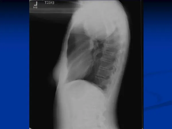
PA and lateral chest films show an anterior mediastinal mass and a large right pleural effusion.
Two contiguous slices from an enhanced chest CT exam show a homogenous, solid, anterior mediastinal mass and a large right pleural effusion. Dx-Lymphoma, Non-Hodgkin, Anterior Mediastinal
PA and lateral chest films show a soft tissue mass in the right posterior costophrenic sulcus.
PA and lateral chest films from the day of admission demonstrate a large round opacity in the left lower lobe that abuts the diaphragm
Two coronal T1 weighted images and one axial T2 weighted image from an MRI exam from the 5th hospital day demonstrate a posterior mediastinal mass that extends into the retrocrural regions of the chest bilaterally and that enhances uniformly. There is no evidence of metastatic disease. Dx-Sequestration, Extralobar
Bone window images from a chest CT exam from the day of diagnosis demonstrate a large spherical calcified left paravertebral mass measuring 12 x 11 x 8 cm in size. There is a pleural effusion and a shift of mediastinal structures to the right. The mass appears to extend via the retrocrural space into the abdomen causing displacement of the left kidney and inferior vena cava. The mass crosses the midline. Some minimal thoracic vertebral body remodeling and rib thinning is seen on the left. No spinal canal invasion or liver metastases are seen
MRI exam performed 3 weeks after diagnosis. Coronal and sagittal T1 weighted images without contrast, and coronal and axial T2 weighted MRI images could not definitely identify the left adrenal gland, and therefore suggested it could be the origin of the midline mass. There was evidence of tumor invasion into several neural foramina and the spinal canal. Dx-Neuroblastoma
Germ Cell • . Almost all of them originate in the anterior mediastinum within or in close contact with the thymus. There is a variety of benign and malignant germ cell neoplasms. The majority of germ cell neoplasms (60–70%) are benign including mostly mediastinal teratoma and dermoid cysts that occur with equal frequency in males and females. • On CT scans, the tumour is heterogeneous and limited with well-defined margins. Dermoid cysts and teratomas contain areas of different densities including fat, soft tissue and cystic • Fatty and cystic components are present in about half of the cases. Occasionally a fat–fluid level may be present and is highly suggestive of the diagnosis. Curvilinear, spherical or irregular calcifications within the mass may be seen Identification of a tooth, while rare, is diagnostic.
Germ Cell Tumors • Malignant germ-cell neoplasms have a male predominance. They include mediastinal seminoma, mediastinal choriocarcinoma, embryonal cell carcinoma, yolk sac tumour and teratocarcinomas • On the radiograph, the mass is similar to benign germ cell neoplasm excepted that the mass itself is often lobular in outline. Metastases may be seen in the lung, pleura or bone. CT or MR features of the tumour are similar to other primary malignant tumours arising within the anterior mediastinum • The mass is lobular and asymmetrical. The margins may be well-defined or irregular. The adjacent mediastinal fat planes may be obliterated, although this is not a feature of definite invasion. The tumour may appear as either a homogeneous soft tissue mass or a heterogeneous mass containing areas of contrast enhancement interspersed with areas of decreased attenuation due to necrosis or haemorrhage • Calcifications are uncommon. • Mediastinal lymphadenopathy may be present
Lymphoma • primary malignant neoplasm of the lymphoreticular system, particularly of the lymphocytes and histiocytes and the derivatives of these two cell types, surrounded by non-neoplastic inflammatory cells. Lymphomas include Hodgkins disease and non Hodgkins lymphoma. Both frequently involve the chest.
Thymoma, • neoplasm arising from thymic epithelium. It is the most common cause of a thymic mass. It presents as an anterior mediastinal mass. • Thymomas occur usually between the ages of 40 and 60 years old, in males or females equally. They are very unusual in patients under the age of 20. Thymomas generally occur as incidental findings discovered on a chest radiograph in otherwise healthy individuals. They may also occur in association with other abnormalities such as myasthenia gravis, red cell aplasia and hypogammaglobulinaemia. Myasthenia gravis, the most frequent association of the three, is present in roughly 50% of patients with thymoma. Approximately 15% of patients with myasthenia gravis have a thymoma • On the chest radiograph, thymomas are depicted as a round or lobulated mass located in the anterior mediastinum • . On lateral films, they often appear as a well-defined mass in the normally clear restrosternal space. • Sometimes, the tumour is situated more inferiorly adjacent to the left or right borders of the heart and occasionally as low as the cardiophrenic angle. • Occasionally the tumour is too small (1 cm) to be depicted on the chest radiograph, and is only detected on CT scans Punctuate or curvilinear calcifications may be seen in both benign or invasive thymomas. • On CT scans, benign thymomas appear as a round or oval mass located in the prevascular space of the mediastinum, or at any level from the thoracic inlet to the diaphragm within the anterior mediastinum. Intratumoral calcifications are present in 20 – 30% of the cases and areas of cystic degeneration are common • Invasive thymomas typically appear as irregular masses growing along pleural surfaces.