Download
1 / 37
370 likes | 377 Views
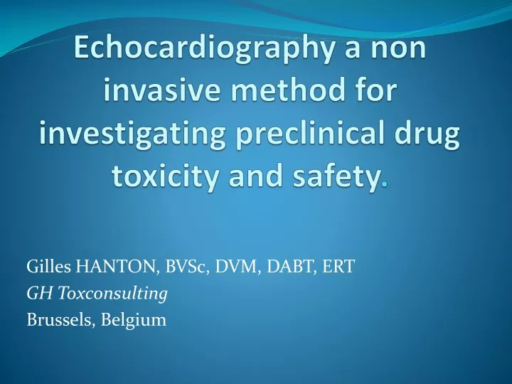
Echocardiography a non invasive method for investigating preclinical drug toxicity and safety. Gilles HANTON, BVSc, DVM, DABT, ERT GH Toxconsulting Brussels, Belgium. What is echocardiography (EC). Ultrasounds (US) are emitted by a transducer

E N D
Echocardiography a non invasive method for investigating preclinical drug toxicity and safety. Gilles HANTON, BVSc, DVM, DABT, ERT GH Toxconsulting Brussels, Belgium
What is echocardiography (EC) • Ultrasounds (US) are emitted by a transducer • Reflection of US on tissues depends on their physical properties (echogenicity) • strong echogenicity : bones, air • weak echogenicity: liquids (blood, urine) • Reception of reflected US by the transducer • Processing of the information and image on the screen
Bidimensional echocardiography (2-D EC) in right parasternal incidenceVisualisation of the heart structures in the plane of the ultrasounds beam: longitudinal section
M-mode echocardiography Positioning of a guidance line through the cardiac structures in 2-D Visualisation of the movements of the cardiac structures
M-mode echocardiographySchematic representation showing measured parameters
The different modesDoppler Assessment of • Quantitative parameters of cardiovascular function • Flows: Stroke volume, cardiac or extra cardiac shunt flow, left coronary blood flow, • Pressure changes across valves and orifices or in cardiac chamber and great vessels • Qualitative blood flow changes: • Laminar vs disturbed flow patterns
Doppler recording of intra-cardiac flows • Visualisation of the heart structures in a 2-D mode section using apical incidence • Positioning of the Doppler Window at the level of the aorta, pulmonary artery, mitral or tricuspid valves • Recording of the changes in blood velocity over a few beats
The different modes: Doppler Four cavities view in apical incidence ( marmosets)
Echocardiography in marmosets: mitral flow Spectrum of distribution of erythrocytes velocities E wave Ventricle diastole A wave Atrial systole
Schematic representation of measurements on a Doppler velocity spectrum of the mitral flow
Pulmonary flow recording (marmoset) Aortic flow recording (marmoset) • Measurements • Vmax, VTI, • Ejection time (ET): from the onset (b) to the end (d) of the velocity spectrum) • Pre-ejection time from the Q wave • of the ECG (a) to the onset of the Doppler velocity spectrum (b), • Acceleration time from the onset to the peak of the velocity spectrum (c) and (d). b d a c
Doppler EchocardiographyCalculated parameters • From tricuspid and mitral flowRatio A/E waves for peak velocity or velocity-time integral : • Relative contribution of atrial systole vs ventricle diastole to ventricle filling • From aortic flowStroke volume = VTI x AA with VTI: velocity time integralAA: aortic diameter measured from M-mode trace
Application of echocardiography in preclinical safety assessment (1) CONSEQUENCES of Cardiac toxicity • Evaluation of morphological changes induced by test compounds (cardiac hypertrophy, dilation…) • Measurement of functional consequences (changes in haemodynamic parameters and in contractility) of compound-induced cardiac lesions • Measurement of haemodynamic changes associated with arrhythmias
Application of echocardiography in preclinical safety assessment (2) CAUSE and MECHANISM of Cardiac toxicity • Evaluation of pharmacological effects of cardiovascular drugs. • Measure of changes in cardiac contractility and in haemodynamic parameters • Clarification of the pathogenesis of cardiac lesions linked to exaggerated pharmacological effects: example of minoxidil
Value of echocardiography in toxicology as a method of refinement • Non-invasive technique- No surgery - No pain or distress for the animal- Only a gentle restraint is needed • No interference on cardiac function: measurement in normal situation • No interference with the measurement of other parameters • No influence on the results of the toxicity study • No medication • No effects of echography on the health status of the animal • Measurements are easily repeatable and allow subsequent follow-up in the same animal
Minoxidil • Potent vasodilator • Cardiac toxicity of minoxidil in the dog • Produces necrosis of left ventricle at suprapharmacological doses (0.5-3 mg/kg) • Is due to the vasodilatory properties of the drug
Example of minoxidilExperimental procedure • Treatment with 0.5 or 2 mg/kg (single dose) • 3 dogs/dose • Measurement of echocardiographic parameters in M-mode and Doppler at different time points before and after dose
Minoxidil effects onparameters of left ventricle function evaluated by M-mode echocardiographyChange (%) in mean values recorded 1 hour after treatment compared to values recorded the day before treatment • PST: Percent of septum thickening; PWT: Percent of left ventricle posterior wall thickening; HR: heart rate • EDV, ESV: end diastolic, end systolic volumes; EF: Ejection fraction
Minoxidil effects on aortic flow measured by Doppler echocardiographyChange (%) in mean values recorded 1 hour after treatment compared to values recorded the day before treatment Vmax: maximum velocity of the wave VTI: velocity time integral ET: ejection time
Minoxidil effects on parameters of left ventricle function evaluated by echocardiography • Increase in contractility • Increase in ejection fraction and percent thickening of the left ventricle wall and septum • Decrease in end systolic volume • Increase in Vmax of aortic flow • Mild increase VTI and consequently in stroke volume • Marked tachycardia leading to • Decrease in ejection time • Decrease in end diastolic volume indicating decreased filling of the ventricle (decrease in inter-systolic time) • Marked increase in cardiac output • Due mainly to tachycardia and to a lesser extent to increase in SV
Relationship between changes produced by minoxidil on cardiac function and the development of cardiac lesions Minoxidil Vasodilation Hypotension Decrease in coronary blood flow Reflex cardiac stimulation Increase in Heart rate Decrease in afterload Decrease in ventricular filling time Increase in CO Increase in ventricular contractility Decrease in EDV and ET Decrease in ESV Increases in PST, PWT, EF , Vmax of Doppler aortic velocity spectrum Increase in oxygen demand in the myocardium Hypoxia in the left ventricle Necrotic lesion
Conclusion of minoxidil study • Echocardiography allows the non-invasive investigation of changes in the cardiac function produced by a vasodilator known to play a critical role in the pathogenesis of cardiac lesions. • In the past, these functional changes were assessed using highly invasive methods
CONCLUSION Echocardiography has potentially a great value as a method for investigation of cardiovascular effects of drugs in toxicology and safety pharmacology
Acknowledgments Establisment echocardiography in dogs and marmosets Drs Pierre Bonnet and Véronique Eder Hopital Bretonneau / University of Tours, France Minoxidil study, Scientific collaboration M. Gautier, PhD studentTechnical collaboration of H. Petinay, N. Mauclair and O. ChristinPfizer Research Center, Amboise, France
Echocardiography in toxicology References • G.Hanton., B; Geffray., A. Lodola.Echocardiography, a non-invasive method for the investigation of heart morphology and function in laboratory dogs: 1. Establishment of the method and reference values for M-mode parameters.Laboratory animals, 32, 173-182, 1998 • G. Hanton, A Lodola.Echocardiography, a non-invasive method for the investigation of heart morphology and function in laboratory dogs: 2. Effects of minoxidil and quinidine on the left ventricle function Laboratory animals, 32, 183-190, 1998 • G. Hanton, Baneux PJREchocardiography in laboratory dogs: a method of refinement for the assessment of cardiovascular toxicology. Example of minoxidil and quinidine.In: Progress in the Reduction, Refinement and Replacement of Animal Experimentation. M. Balls, A.-M. van Zeller and M. E. Halder editors, Elsevier, Amsterdam, 2000, pp 1175-1186 • G. Hanton, Gautier M., Bonnet. P.Using M -mode and Doppler echocardiography to investigate the cardiotoxicity of minoxidil in Beagle dogs.Arch. Toxicol, 78, 40-48, 2004 • G.Hanton , Gautier M., Herbet A., Bonnet P.Effect of milrinone on echocardiographic parameters after single dose in Beagle dogs and relationship with drug-induced cardiotoxicity.Toxicol Letters, 155, 307-317, 2005 • Serriere S., Tranquart F., Hanton G.Sonographic exploration of the mesenteric and renal arterial blood flows in adult rats. ToxicolLett., 158, suppl 1, S237, 2005 • Boissiere J, Gautier M, Machet M-C, Hanton G, Bonnet P, Eder V.Doppler tissue imaging in assessment of pulmonary hypertension-induced right ventricle dysfunction. Am. J. Physiol: Heart Circ. Physiol., 269, H2450-H2455, 2005 • Serrière S., Tranquart F., Hanton G.Echographic recording of uterine, umbilical and fetal cerebral blood flow in pregnant rats.Toxicol Letters, 164S, S306, 2006 • G. Hanton., Eder V. Bonnet P., Rochefort G.Y. Echocardiography in marmosets: a non-invasive method for the assessment of cardiovascular toxicology and pharmacology. In: GF Weinbauer, F Vogel (eds). NovelapproachestowardsprimatetoxicologyWaxmann Publishing Co. Münster/New York, 2006 • G. Hanton, Eder V., Rochefort G., Bonnet P., Hyvelin JM.Echocardiography, a non-invasive method for the assessment of cardiac function and morphology in pre-clinical drug toxicology and safety pharmacology. Exp. OpinMetabol. Toxicol., 4 (6), 2008 Footer
THANK YOU for your attention Dr. Gilles Hanton GH Toxconsulting Brussels, Belgium gilles.hanton@yahoo.fr
2-D echocardiography in right parasternal incidence Scanning in transverse section
M-mode echocardiography of the upper part of the heart in a marmoset. The guidance line is positioned across the aorta and left atrium .The movements of aorta (AO) and left atrium (LA) marmoset, are recorded over time.
Color Doppler of intra-cardiac flows: ventricular diastole (marmoset). The flow from left atrium to left ventricle trough mitral valves appears in red.
Color Doppler of intra-cardiac flows: ventricular systole (marmoset) The aortic flow appears in blue