Download

1 / 54
870 likes | 1.94k Views
CSOM ATTICO ANTRAL DISEASE. DR MUBEENA. CHOLESTEATOMA. The presence of stratified squamous epithelium in the middle ear or mastoid. Skin in the wrong place. Misnomer ?. “ Cholesteatoma " coined by the German physiologist Johannes Müller in 1838, is a misnomer

E N D
CSOMATTICO ANTRAL DISEASE DR MUBEENA
The presence of stratified squamous epithelium in the middle ear or mastoid. • Skin in the wrong place.
Misnomer ? • “Cholesteatoma" coined by the German physiologist Johannes Müller in 1838, is a misnomer because this entity does not contain cholesterol • the white-yellow keratin flakes found within cholesteatomas grossly resemble cholesterol crystals • ‘oma’ is a suffix for tumour
SYNONYMS:- • KERATOMA • EPIDERMOSIS • CHOLESTEATOSIS
Choleasteatoma consists of : • PERIMATRIX – stroma of connective tissue • MATRIX – keratinising squamous epithelium resting on thin stroma of fibrous tissues . • KERATIN DEBRIS – central white mass produced by the matrix
Cholesteatoma has the capacity for progressive and independent growth at the expense of underlying bone and has a tendency to recur after removal.”
CLASSIFICATION:- • CONGENITAL • ACQUIRED- primary acquired secondary acquired
Pathogenesis • Congenital • Arise from embryonal rests of epithelial cells • COMMON SITES- middle ear – ASQ commonest site mastoid cerbellopontine angle petrous apex
CONGENITAL CHOLESTEATOMAS • Most involve the anterior superior quadrant of the middle ear
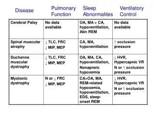
Clinical symptoms • Conductive hearing loss. • Otalgia • Vertigo or SNHL if labyrinth is invaded . IMAGING HRCT CT Scan – confirm location and size
LEVENSON’S CRITERIA • White mass behind the normal ear drum • • Normal pars flaccida and pars tensa • • No prior history of perforation / Otorrhoea • • No previous otological procedures
Surgical management of a congenital cholesteatoma • complete removal of the cholesteatoma matrix • The isolated middle ear cholesteatoma • removed transtympanically • Routine middle ear reconstructive techniques may be used if ossicles are eroded or removed or if the tympanic membrane is sacrificed.
The pathogenesis of acquiredcholesteatoma There are four basic theories : • Invagination of the tympanic membrane • (retraction pocket cholesteatoma) (2)basal cell hyperplasia (3) epithelial ingrowth through a perforation (the migration theory) (4)squamous metaplasia of middle ear epithelium
Invagination theory- Wittmack 1939 Eustachian tube dysfunction -negative ME pressure -pars flaccida retraction -squamous debris collected in the sac
2. Epithelial invasion theory: Haberman 1889:- keratinising squamous epithelium from the surface of the TM or meatus invades into the middle ear from perforation in TM.
3. Basal cell hyperplasia theory: Reudi’s theory • The basal cells of germinal layer of skin proliferate under the influence of infection and lay down keratinising squamous epithelium
4. Squamous metaplasia theory: Wendt 1873:- Simple cuboidal epithelium of ME cleft could undergo metaplastic transformation into keratinising epithelium because of repeated infection.
Primary acquired cholesteatoma • Called primary as there is no history of previous perforation • Secondary to ET dysfunction • Retraction pocket choleasteatoma in Pars flaccida or PSQ retraction pocket • Invagination theory is most accepted • Keratin debris collects in the retraction pocket
Primary acquired cholesteatoma Normal TM Mesotympanic cholesteatoma primary acquired cholesteatoma
WHY ATTIC AND PSQ IS PREDISPOSED FOR RETRACTION ? • There are a fewer connective tissue fibres within the lamina propria (middle layer of TM) as compared to pars tensa • Normal migratory pattern of TM epithelium changes within retraction pocket . This results in keratin formation and choleasteatoma • PSQ – weakest quadrant of the pars tensa
Secondary Acquired Cholesteatoma • Migration Theory – most accepted • Originates from a tympanic membrane perforation • As the edges of the TM try to heal, the squamous epithelium migrates into the middle ear
Microbiology:- Pseudomonas Proteus Mixed aerobic and anaerobic organisms
Behaviour of choleasteatoma BONE RESORPTION • Pressure necrosistheory- unlikely • Enzymatic theory by cytokine mediated inflammation : Inflammation Release IL-1,IL-6, IL-11, TGF-alpha cause osteoclast activation Acid phosphatase, collagenase, acid proteases, cathepsin like proteolytic enzyme, matrix metalloproteases
bone erosion • Ossicles : most long process of incus • Bony labyrinth • facial nerve canal • tegmen tympani • Sinus plate
ATTICOANTRAL TYPE • It involves POSTERIOR SUPERIOR part of the middle ear cleft (attic, antrum , posterior tympanum and mastoid)
PATHOLOGY • Cholesteatoma • Osteitis and granulation tissue • Ossicular necrosis • Cholesterol granuloma
OSSICULAR NECROSIS • LONG PROCESS OF INCUS • Hearing loss more than TTD • Cholesteatoma hearers
CHOLESTEROL GRANULOMA • Granulation tissue with foreign body giant cells surrounding cholesterol crystals • Reaction to longstanding retention of secretion or haemorrhage
Symptoms • Ear discharge • Impaired hearing- cholesteatoma hearer • Tinnitus • Vertigo
Signs • Retraction pocket in early stage • Characteristic discharge • ‘Fishy odor of cholesteatoma • Attic/ marginal or total perforation • Cholesteatoma seen as • Pearly white sac through translucent TM or as • Pearly white flakes of epithelium through mouth of the sac, or as • In-growing skin though perforation
Other signs • Granulation tissue in the attic/ postero-superior quadrant • Attic crust • Vascular polyp • Attic widening
Evaluation • Examination under microscope • Cultures should be obtained in infected ears • X RAY mastoid– extent of bone destruction Cholesteatoma- sclerosis with cavity. • Pure tone audiometry • HIGH RESOLUTION CT SCAN OF TEMPORAL BONE
Evaluation 1. Examination under microscope • Retraction pocket • Choleasteatoma- suction clearance and examination • Granulation from diseased bone • Aural polyps • Ossicles
Evaluation • PURE TONE AUDIOMETRY – usually conductive loss, may vary greatly; confirm with tuning forks • pre-operative assessment • degree of hearing loss • Type of hearing loss
Computed Tomography • Erosion of scutum • Destruction of ossicular chain • Erosion of the labyrinth (fistula) • Low tegmen / tegmen defect • Facial nerve dehiscence • Petrous Apex Involvement