Download
1 / 67
700 likes | 1.09k Views
Urinary System Assessment, Diagnostics , Diseases, and Treatments. Skin Turgor-commonly used to assess degree of dehydration or fluid loss. Dehydration. Dry skin Sunken fontanelle No tears Cracked lips Sunken Eyes Headaches Haven’t urinated. Edema.

E N D
Urinary System Assessment, Diagnostics, Diseases, and Treatments
Skin Turgor-commonly used to assess degree of dehydration or fluid loss.
Dehydration • Dry skin • Sunken fontanelle • No tears • Cracked lips • Sunken Eyes • Headaches • Haven’t urinated
Edema • Swelling caused by fluid retention- excess fluid is trapped in the body's tissues. • Commonly occurs in the hands, arms, ankles, legs and feet.
Edema may be generalized or local. It can appear suddenly, but usually develops subtly - the patient may first gain weight, or wake up with puffy eyes. Many patients wait until symptoms are well advanced before seeking medical help.
Daily Weights • Body weight change provides the simplest and most accurate index of hydration status.
Bladder Distention • Urinary retention is the inability to voluntarily void urine. • Urinary retention can lead to bladder distention.
Bladder Scanner • A noninvasive method of assessing bladder volume and other bladder conditions using ultrasonography to determine the amount of urine retention or post-void residual urine.
Strict I & O (in a 24 hour period) • Intake: fluids, food, IV fluids, blood products, medications. • Output: urine, vomit, drains, feces, ostomies etc. • Always measured in ml • Monitor optimal fluid balance.
Renal system (physical assessment) • General state of health- fatigue, lethargy, & diminished alertness. Nocturia, dysuria, hesitancy, dribbling, hematuria, polyuria, etc. • Palpation- No costovertebral angle tenderness, nonpalpable kidney & bladder, no palpable masses. • Percussion: Tenderness in the flank may be detected by fist percussion. If CVA tenderness & pain are present, indicate a kidney infection or polycystic kidney disease • Auscultation: The abdominal aorta & renal arteries are auscultated for a bruit, which indicates impaired blood flow to the kidneys
Flank Pain • The costovertebral angle, sometimes referred to as CVA, is the space created by the downward lateral slop of the last rib and the beginning of the lumbar vertebrae or back bones of the spine.
Urine Output • The expected urine output for an adult is 0.5ml/kg/hr. This roughly equates to 30-40ml per hour in an average sized adult. Children should have 1ml/kg/hr of urine output.
Lab Tests • BUN (blood urea nitrogen): Measurement of urea in blood. Normally low because urea is excreted in the urine. • Creat (Creatinine): The substance creatine is formed when food is changed into energy through a process called metabolism. Creatine is broken down into another substance called creatinine, which is taken out of your blood by the kidneys and then passed out of your body in urine. High levels in the blood indicate kidney problems.
Renal Systems (Diagnostic test) • Urinalysis-evaluation of the renal system & for determining renal disease. • Wash perineal area & use a clean container. • Obtain 10 to 15 mL of the 1st AM sample • If the client is menstruating, indicate this on the lab. requisition form. • Specific Gravity-measures the kidney’s ability to concentrate urine. Measured by multiple-test dipstick (most common method). • A decrease in SG (less conc. urine) occurs with increased fluid intake, diuretic administration, diabetes insipidus. • An increase SG (more conc. Urine) occurs with insufficient fluid intake, decreased renal perfusion, or the presence of ADH.
Renal Systems (Diagnostic test) • Urine Culture & Sensitivity- identifies the presence of microorganisms & determines the specific antibiotics that will treat the existing microorganisms. Note that urine from a client who forced fluids may be too dilute to provide a positive culture.
Renal Systems (Diagnostic test) • Creatinine clearance test- A blood & timed urine specimen that evaluates kidney function. • Blood is drawn at the start of the test & the AM of the day that the 24-hour urine specimen collection is complete. Maintain the urine specimen on ice or refrigerate.
Renal Systems (Diagnostic test) • Uric acid- A 24-hour collection to diagnose gout & kidney disease. • Encourage fluids & a regular diet during testing. Place the specimen on ice or refrigerate.
KUB (Kidney, ureters, bladder) radiograph-An x-ray film that views the urinary system & adjacent structures; used to detect urinary calculi.
Renal Systems (Diagnostic test) • Intravenous pyelogram (IVP)- the injection of a radiopaque dye that outlines the renal system. Performed to identify abnormalities in the system. Withhold food & fluids after midnight before the test. Inform the client abt. Possible throat irritation, flushing of the face, warmth or salty taste that may experienced during the test.
Renal Systems (Diagnostic test) • Renal angiography- the injection of a radiopaque dye through a catheter for examination of the renal arterial supply. Assess the client for allergies to iodine, seafood & radiopaque dyes. Inform about possible burning feeling of heat along the vessel when the dye is injected.
Renal Systems (Diagnostic test) • Cystoscopy & Biopsy- the bladder mucosa is examined for inflammation, calculi or tumors by means of a cystoscope, a biopsy may be obtained.
Urinary Tract Infection (UTI) • Inflammation of the bladder from infection or obstruction of the urethra. • The most common causative organism are E. coli, Enterobacter, pseudomonas, & serratia. • More common in women because they have shorter urethra than men, & the location of the urethra in women is close to the rectum. • Sexually active & pregnant women are most vulnerable to UTI.
Urinary Tract Infection (UTI) • Causes: Allergens or irritants, such as soaps, sprays, bubbles bath • Bladder distention, calculus, hormonal changes influencing alterations in vaginal flora. • Indwelling urethral catheter, loss of bacterial properties of prostatic secretions in the male • Sexual intercourse, urinary stasis, use of spermicides, wet bathing suits
Urinary Tract Infection (UTI) • Assessment: Frequency & urgency, burning on urination, voiding in small amount, inability to void, incomplete emptying of the bladder, lower abdominal discomfort or back discomfort, cloudy, dark, foul smelling urine, hematuria, bladder spasms, malaise, chills, fever, nausea & vomiting. • Implementation: Obtain urine C/S to identify bacterial growth. Instruct to force fluids up to 3000 mL a day. Provide meticulous perineal care with an indwelling catheter. Instruct to avoid alcohol. Provide heat to abdomen or sitz bath for complaints of discomfort
Urinary Tract Infection (UTI) • Provide relief by administering analgesics such as Pyridium or combination agents (Urised). Alert that urine color will be orange & blue or green with combination agents. • Teach the use of nonpharmacologic technique- heating pad, warm showers. • Treat with antibiotics.
Urinary Tract Infection (UTI) • Impaired urinary elimination r/t UTI as manifested by bothersome urgency, hematuria or concern over altered elimination pattern • Obtain midstream voided specimen for C/S. • Administer antimicrobial drugs. • Teach signs & symptoms of UTI. • Encourage adequate fluid to help prevent infection and dehydration.
Urinary Tract Infection (UTI) • Teaching: teach good perineal care & to wipe from front to back. • Instruct to void every 2 to 3 hours. • Instruct to void & drink a glass of water after intercourse. • Encourage menopausal women to use estrogen vaginal creams to restore pH. • Instruct the female to use water- soluble lubricants for coitus, especially after menopause.
Foley Catheter • A flexible tube that is often passed through the urethra and into the bladder. The tube has two separated channels, or lumens, running down its length. One lumen is open at both ends, and allows urine to drain out into a collection bag. The other lumen has a valve on the outside end and connects to a balloon at the tip; the balloon is inflated with sterile water when it lies inside the bladder, in order to stop it from slipping out. Foley catheters are commonly made from silicone rubber or natural rubber.
Indwelling urinary catheterization is usually performed to monitor the urine output of individuals undergoing surgery or after trauma or obstruction (of the urinary tract or to drain the bladder in individuals finding it difficult to void (frequently during serious illnesses).
Straight Cath (In and out cath) • A straight catheterization may be performed to obtain a sample of urine for laboratory analysis. Urinary catheterization may be performed in individuals who have urinary incontinence or who cannot empty their bladder.
Urostomy-A diversion of the urinary flow away from the bladder, resulting in output through the abdominal wall.
Nephrostomy-small rubber tube that is placed through a hole in the skin and that extends into the kidney.
Urolithiasis • Formation of urinary stones; urinary calculi formed in the ureters. • When a calculus occludes the ureter & blocks the flow of urine, the ureter dilates, producing a condition known as hydroureter. • If the obstruction is not removed, urinary stasis results in infection, impairment of renal function on the side of the blockage, & resultant hydronephrosis & irreversible kidney damage.
Urolithiasis • Causes: Family history of stone formation • Diet high in CA, vitamin D, milk, protein, purines • Obstruction & urinary stasis • Dehydration • Use of diuretics, which can cause volume depletion • Immobilization • Hypercalcemia, & hyperparathyroidism • Elevated uric acid, such as gout
Urolithiasis • Assessment: Nausea, vomiting, dietary intake of purines, phosphates, low fluid intake; chills. • Elimination: Decreased u/o, urinary urgency, feeling of bladder fullness. • General: Acute, severe colicky pain in flank, back, abdomen groin or genitalia; burning sensation on urination, dysuria,anxiety. • Skin: warm, flushed skin or pallor with cool. • Urinary: tenderness on palpation on renal areas, passage of stone(s). • Increased BUN & creatinine; WBC, calcium, phosphorus, uric acid. • KUB- calculi or anatomic changes on IVP
Urolithiasis • Treatment: Force fluids up to 3000 mL/day, unless contraindicated-to facilitate the passage of the stone & prevent infection. • Strain all urine for the presence of stones. • Turn and reposition immobilized clients. • Administer analgesics & response to pain. • Instruct in the diet specific to the stone composition.
Urolithiasis • Surgical therapy: • Nephrolithomy- incision into the kidney to remove the stone. • Pyelolithotomy- incision into the renal pelvis to remove the stone. • Ureterolithotomy-removal of stone in the ureter. • Cystotomy- indicated for bladder calculi. • Lithotripsy- procedure used to eliminate calculi in the kidney. Hematuria is common after the procedure. A stent is often placed after the procedure to promote passage and to prevent obstruction, then removed 1 to 2 weeks after lithotripsy.
Nephrolithiasis is kidney stones (renal calculi). Usually composed of uric acid or calcium salts.
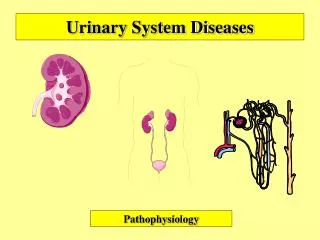
Glomerulonephritis: Inflammation of the kidney glomerulus. • Cystitis: Inflammation of the bladder. • Pyelonephritis: Inflammation of the renal pelvis and renal medulla. • Interstitial Nephritis: Inflammation of the connective tissue that lies between the renal tubules.
Renal Tumors • May be benign or malignant: Common sites of metastasis include bone, lungs, liver, spleen or other kidney. • Assessment: Dull flank pain, palpable renal mass, painless hematuria. Unknown cause. • Treatment: Radical nephrectomy: Removal of the entire kidney, adjacent adrenal gland & renal artery & vein. • Radiation therapy & chemotherapy.
Renal Tumors • Implementation: Monitor abdomen for distention caused by bleeding • Observe bed linens under the client for bleeding • Monitor for hypotension, decreases in urinary output & alterations in LOC, indicating hemorrhage. • Monitor urinary output • Do not irrigate or manipulate the nephrostomy tube if in place.
Acute renal failure (also called acute kidney injury) means that your kidneys have suddenly stopped working. Your kidneys remove waste products and help balance water and salt and other minerals (electrolytes) in your blood. When your kidneys stop working, waste products, fluids, and electrolytes build up in your body. This can cause problems that can be deadly.
ARF • Symptoms of acute renal failure may include: • Little or no urine when you urinate. • Swelling, especially in your legs and feet. • Not feeling like eating. • Nausea and vomiting. • Feeling confused, anxious and restless, or sleepy. • Pain in the back just below the rib cage. This is called flank pain.