Download
1 / 47
500 likes | 709 Views
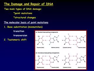
ROS, RNI, DNA Damage and Repair Signaling. Lynn Harrison, Ph.D. Department of Molecular and Cellular Physiology. Endogenous Cellular Factors That Damage DNA. Replication errors e.g. imbalance in the nucleotide pools result in mismatch DNA instability deamination of bases

E N D
ROS, RNI, DNA Damageand Repair Signaling Lynn Harrison, Ph.D. Department of Molecular and Cellular Physiology
Endogenous Cellular Factors That Damage DNA • Replication errors • e.g. imbalance in the nucleotide pools • result in mismatch • DNA instability • deamination of bases • depurination/depyrimidination of DNA • loss of base • Reactive oxygen and nitrogen species • DNA oxidation and deamination
Types of DNA Damage • Base Damage • Loss of bases - abasic or apurinic sites • Strand breakage • Protein-DNA cross-links • DNA-DNA cross-links
Reactive oxygen species • Produced by normal cellular metabolism • Mitochondria utilize ~85% O2 in cell and are a major source of ROS • Damage DNA, protein and lipid. • Some forms in cell are: • Hydrogen peroxide (H2O2) • Superoxide radical (O2•-) • Nitric oxide (•NO) • Hydroxyl radical (HO•)
Fenton Reaction • Metal-catalyzed formation of HO• radicals. This reaction can function in a redox cycle in which a transition metal ion transfers electrons from donors. DNA(FeII) + H2O2 DNA(FeIII) + OH + OH-
Hydroxyl radicals • Small modifications to bases, many types/base • Single and double strand breaks • Abasic sites (loss of base) • Approximately 100 different types of damages identified dR Thymine
The Chemistry of Nitric Oxide Dictates its Physiological Activity Metal Complexes/Alkyl Radicals Guanylate Cyclase Cytochromes C,O,N Radicals (Lipid Radicals) Direct NO L-Arginine eNOS nNOS iNOS O2 orO2- Indirect RNOS Oxidation Nitration Nitrosation DNA Strand Breaks Lipid Peroxidation Hydroxylation Nitrotyrosine Nitroguanosine Nitrosothiols Nitrosamines
O O H C H C 3 3 N N N N H C H C 2 3 O H O N H N N H N N 2 Nitrosamine-Mediated Alkylation of DNA Bases Hydroxylation P450 N-Nitrosodimethylamine CH2O + H2O CH3N2+ + C H O 3 N H N N H N N 2 O6Methylguanine Guanine
Nitrosative Deamination of DNA Bases by NO-Derived N2O3 ArNHNO ArNH2 +N2O3 ArNHNO + H+ ArN2+ +H2O ArOH + N2 +H+ ArN2+ +H2O ArNH2represents DNA bases containing an exocyclic amino group: Cytosine, methylCytosine, Guanine or Adenine
Guanine Biological Consequences of DNA Damage • Block to DNA replication • e.g. thymine glycol, abasic site • Mutagenesis • e.g. 8-oxoguanine pairs with A as well as C dR
Deletions • Common with ionizing radiation • Due to strand breakage • Chromosomal aberrations • Double strand breaks on different chromosomes or chromatids • Aberrant transcription • Breakage or abasic sites believed to result in reduced transcription • Gaps in DNA result in deleted transcripts • Consequences are altered/ mutated proteins
Types of Repair • Mismatch Repair • Repairs mismatches, these can be generated by replication • Nucleotide excision repair • Repairs bulky lesions predominantly, also some small oxidative lesions • Base excision repair • Repairs small lesions e.g. oxidative damage and deaminations • Non-homologous end-joining • Repairs double-strand breaks in all phases of the cell cycle • Homologous recombination • Repairs double strand breaks in the S or early G2 phases of the cell cycle
What activates the “alarm” signals after DNA damage • Repair proteins that initiate the different pathways recognize lesions specifically. • Believed to be constantly “patrolling” the DNA for damage, since damage is constantly occurring. • If repair cannot handle the damage then the cell cycle checkpoints and possibly apoptosis are activated. • Exception maybe the generation of a DSB where signaling is rapid. Activation may occur at a similar time as repair.
e.g. BER RPA Cell cycle checkpoint Initiation of signaling ROS, RNI • ATM and ATR are members of the PI-3-kinase-like family of kinases. • Much of the signaling is through protein phosphorylation Sustained ssDNA Jeggo & Lobrich Radiation Protection Dosimetry 2007
Activation of ATR ROS, RNI Cell cycle checkpoint Jeggo & Lobrich Radiation Protection Dosimetry 2007 Occurs in S phase due to replication block Or when there is excessive ssDNA
Not generate ssDNA ATR signaling due to RPA bound to ssDNA caused by stalled replication or blocking lesion DNA Repair 6, 953 • MCM2-7 • Replicative DNA helicase, unwinds the DNA • Cdc45 • Essential for initiation and elongation of DNA synthesis • Pol a, d and e • DNA polymerases • PCNA • Proliferating cell nuclear antigen • RPA • Replication protein A, binds to ssDNA
Rad9-Hus1-Rad1 (Binds to TopBP1, Required for activation of ATR kinase) ATR + ATRIP (P by ATR, essential for activity) Rad17 (P by ATR, aids loading 911 binds to claspin) TopBP1 (9-1-1 loads TopBP1 + activates ATR) P P P Assembly on ssDNA RPA on SSDNA Claspin (P by ATR Needs Rad17)
ATR + ATRIP P Chk1 P P P TopBP1 Claspin After loading, Chk1 that is associated with the chromatin is phosphorylated • Claspin channels ATR to phosphorylate Chk1 • TopB1 assists ATR-ATRIP in phosphorylating numerous substrates including Chk1 Rad9-Hus1-Rad1 Rad17 P
Cdc25 phosphorylase Wee1 kinase ATR phosphorylates Chk1 to stop progression to mitosis • DNA damage causes blockage of cell cycle Chk1 Chk1 Chk1 Chk1 Chk1 Chk1 Chk1 Chk1 Chk1 Chk1 Chk1 Chk1 P P Progression S→M cytoplasm Adapted from Current Biology 16, 150
Results of Activation of ATR and Chk1 DNA Repair 6, 953
Summary S phase damage DNA Repair 6, 953
Most DSBs are repaired by homologous recombination in S phase Mre11/Rad50/Nbs1 RPA binding Mutation Research 614, 95
Activation of ATM and DSB repair ROS, RNI Jeggo & Lobrich Radiation Protection Dosimetry 2007 Occurs when there is a double strand break Can be cross-talk between the two kinases ATM and ATR
Mammalian cells End-binding and synapsis Ku70, Ku80 DNA PKcs Terminal processing PNK 3’ P’ase Artemis Fen1 WRN, BLM BRCA1 Polm (Poll) Prim1 (Pola complex) Ligation DNA Ligase IV XRCC4 Adapted from Wilson et al 2003 TIBS 28,62.
Damage/ repair foci • Occurs if the protein is retained at the site of damage • Can be produced with ionizing radiation either targeted to the nucleus or by irradiating the whole cell • Can also be produced with a laser • After irradiation cells are fixed and immunohistochemistry used to detect proteins • Or can use fluorescent tagged proteins and in the case of the laser the proteins can be watched in real time as they move in the nucleus and redistribute from a diffuse appearance to high intensity foci • Has been used to determine the order of proteins moving to the DNA
Pre 10” 60” 180” Pre 10” 60” 180” Irradiation site Irradiation site YFP-DNA-PKCs YFP-DNA-PKCs NHEJ proteins form foci only if there is a lot of damage Cells contain Ku • Not possible to see Ku proteins well even under these conditions • Likely this is due to the fast on and off movement of the proteins • Note DNA-PKcs is visible within 10 seconds and only when Ku is present in the cell Cells do not contain Ku Data from David Chen, UT Southwestern
ATM activation and NHEJ likely occur at the same time • ATM is activated quickly and is found in foci • But the proteins involved in NHEJ are also activated rapidly but not retained at the damage unless it is severe. • Site of a DSB is marked by a chromatin modification called gamma H2AX • H2AX is a variant of Histone 2A and it is phosphorylated and binds to the DNA near a DSB and spreads along the DNA up to a megabase pair flanking the break site • Believed the resolution of the foci indicates repair
Activation of ATM • ATM is a PI-3 kinase that phosphorylates proteins • Loss of this protein results in radiosensitization • It is required to block the cell cycle in G1, S and G2 • Exists as an inactive dimer, chromatin changes believed to cause activation and it autophosphorylates Nature 421, 499
ATM activation does not require binding to a DSB • Phosphorylation is detectable as soon as cells can be collected after irradiation and is maximal in 5 minutes after 0.5 Gy (~50% of protein is active) • At 0.5 Gy there are only about 18 DSBs in the genome of the cell • Can also be induced by a few restriction site cuts • Can be activated by hypotonic swelling of cells • Can be activated by chemicals known to alter chromatin modifications and packaging e.g. trichostatin A • Induction of breaks thought to cause relaxation of DNA structure and this is sensed by ATM
Breast cancer associated protein Structural maintenance of chromosome family protein • ATM can also be activated by: • Retinoic acid (not cause DNA damage) • MNNG when DNA is not present • 15-deoxy-delta(12,14)-prostaglandin J(2), which modifies SH groups • REDOX activation? 3 1 1 Exo/endonuclease Needed for ATM at DSB 2 Involved in blocking cell cycle in S phase Cold Spring Harbor Symposia on Quantitative Biology, vol LXX, 99-109 Kitagawa & Kastan
-53BP1 and MDC1 are mediators of ATM signaling -They move to the DNA and are phosphorylated by ATM -May be docking proteins for other signaling proteins -No known activity associated but are required for ATM signaling -BRCA1, MDC1 and 53BP1 needed for efficient autophosphorylation of ATM • Phosphorylated ATM is needed for gamma H2AX modification. DNA PK from NHEJ may also do the phosphorylation • Gamma H2AX required for repair foci formation J. Cell Biol. 173,195
Summary of proteins involved in signaling that are retained at damage NHEJ ATM signaling Checkpoint signaling ATR and HR
ATM signaling results in blockage in G1, intra S and G2/M Chk1 Chk2 p53 Smc1 Chk1+Chk2
G1 block by p53 Chk2 p21 Cyclin A and E associates with Cyclin dependent kinase 2 G1 to S progression
Chk1 and Chk2 Chk1 and 2 Phosphorylation of Cdc25A (inactivates phosphatase) ↓ Cdk2-cyclin E Prevents Cdc45 binding to origins of replication Blocks replication and S phase
Chk2 also alters G2/M ATM MDC1 Chk2 Chk1 ↓ Cdc25 phosphorylase ↑ Phosphorylated Cdc2/cyclinB Unable to progress to mitosis
Reactive nitrogen intermediates result in stabilization of p53
Ref1 found to influence the transcriptional activity of p53 • Loss of Ref1 resulted in reduced induction of p21 and BAX • Involved in transcriptional and hence p53 pro-apoptotic actions
ATM implicated in redox control in cells • ATM was found due to a human disease • Ataxia Telangiectasia • 1 per 40,000 live births, autosomal recessive • Cerebellar ataxia- staggering gait, severe muscular uncoordination, progressive mental retardation • Ataxia – blood vessel dilation in the eyes • Increased cancer incidence • Defective immune system • Knockout mice have higher levels of oxidative stress, found higher H2O2, without increase in catalase • Believe cells of CNS die due to this enhanced stress • Mechanism for ATM to control oxidative stress in cells is not known
ATM also implicated in insulin response • AT patients show glucose intolerance and insulin-resistance • Insulin stimulation of cells seen to activate ATM and to release elF-4E allowing increased translation of specific transcripts • ATM in vitro found to phosphorylate elF-4E binding protein 1 • ATM knock-out mice show delayed insulin secretion when posed with a glucose challenge • Hence ATM maybe involved in insulin signaling • and metabolic function • Altering of metabolic function could be a cause of the enhanced oxidative stress in the cells.
Bystander effect • Irradiation of one cell, triggers stress signals in neighboring cells that were not hit by the radiation • Do not need to damage the DNA of the irradiated cell • The bystander cells do get chromosomal aberrations, mutation and it can cause transformation and cell death. • Bystander cells may have DNA damage as signaling pathways are triggered as if DSB induction has occurred. • Cytoplasmic irradiation and mitochondria as well as ROS and RNI have been implicated.