Download

1 / 131
1.38k likes | 2.07k Views
Thalassemia and Hemoglobinopathies. Ahmad Shihada Silmi Msc, FIBMS Staff Specialist in Hematology Medical Technology Department Islamic University of Gaza. Quiz. What is structure of hemoglobin A? What is the normal hemoglobin types in normal adults?

E N D
Thalassemiaand Hemoglobinopathies Ahmad Shihada Silmi Msc, FIBMS Staff Specialist in Hematology Medical Technology Department Islamic University of Gaza
Quiz • What is structure of hemoglobin A? • What is the normal hemoglobin types in normal adults? • Hemoglobin is composed of……….. and……… • There are …. types of globin chains which are…….. • Normally, rate of globin chain production is equal or not equal.
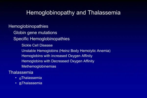
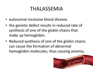
Thalassemia • Syndromes arising form decreased rate or absence of globin chain synthesis. • The resulting imbalance-globin chain synthesis takes place, giving rise to the excess amount of the normally synthesized globin chain.
Hemoglobinopathies • The syndrome arising from the synthesis of abnormal hemoglobin or hemoglobin variants. • Rate of globin chain synthesis are theoritically normal. • Abnormal hemoglobins have different properties from the normal ones.
Incidence of thalassemia in Thailand • a-thalassemia : 20-30 % • b-thalassemia : 3-9 % • Hb E : 8-70 % (very high in E-sarn) • Hb Constant Spring : 1-6 % • Thalassemia disease : 1%
Mode of inheritance • Autosomal recessive • Heterozygote or double heterozygote are not affected. • Homozygote or compound heterozygote are affected.
How to name thalassemia? • Named after globin chain that is abnormally synthesized !!!! • Reduced or absent a-globin chain : a-thalassemia • Reduced or absent b-globin chain : b-thalassemia • Reduced or absent g-globin chain : g-thalassemia • Reduced or absent d-globin chain : d-thalassemia • Reduced or absent gdb-globin chains : gdb-thalassemia
Common types of thalassemia • a-thalassemia • b-thalassemia
α Thalassemia • Absence of α chains will result in increase/ excess of g globin chains during fetal life and excess β globin chains later in postnatal life. • Severity of disease depends on number of genes affected.
SymbolismAlpha Thalassemia • (/): Indicates division between genes inherited from both parents: • / (Normal) • Each chromosome 16 carries 2 genes. Therefore the total complement of genes in an individual is 4.
SymbolismAlpha Thalassemia • (-) : Indicates a gene deletion: • -/ • + Thalassemia (one gene deletion) • 3 functional working genes. • Called thal 2.
SymbolismAlpha Thalassemia • (-) : Indicates a gene deletion: • --/ • 0 Thalassemia (two gene deletion) in the same chromosome. • 2 functional working genes. • Called thal 1.
SymbolismOther Thalassemia • Superscript T denotes nonfunctioning (mutated gene, not deletion) gene: • T
Classification & TerminologyAlphaThalassemia • Normal / • Silent carrier - / • Minor -/- • --/ • Hb H disease --/- • Barts hydrops fetalis --/--
α Thalassemia • Defects in α globin affecting the formation of both fetal and adult hemoglobins, thus, producing intrauterine as well as postnatal disease. Unlike β thalassemia, why??
α Thalassemia • The most common cause of α thalassemia is due to α gene/s deletions. • The most likely mechanism for α gene deletion is due to homologous pairing between α1 and α2 and recombination. This results in loss of α gene. Other causes of α thalassemias are deletions in the locus control regions (HS40) or chain termination mutations (nonsense mutations).
Types of a-thalassemia • a-thalassemia-1 or ao-thalassemia (--) • a-thalassemia-2 or a+-thalassemia (-a) --/aa -a/-a
Compound heterozygotes • Hb H disease ( --/-a) • Hb Bart’s hydrops fetalis syndrome (--/--)
a1 a1 a1 a1 a2 a2 a2 a2 a1 a1 a1 a2 a2 a2 a2 a1 a2 α Thalassemia Four α gene deletions Hydrops fetalis or also called: Erythroblastosis Fetalis. Normal Hb Two α gene deletions α-Thal1 One α gene deletion α-Thal2 Three α gene deletions Hb-H disease
α Thalassemia • As said, the genetic basis of α thal is mostly deletions: If you have 4 functional α genes, then you are normal. • With 3 functional α genes, you are a silent carrier. • With 2 functional α genes you have α thalassemia trait which is clinically benign, but there is mild microcytic anemia. • With only one functional α chain, you have severe hemolytic anemia with primarily HbH, composed of 4 β chains (β4). This is clinically severe. • In the absence of α chain in the fetus, the gamma forms a tetramer of globin chains, and is called Hb Bart’s. • Both Hb-H and Hb-Barts are high affinity Hbs, thus neither of them is capable of releasing oxygen to the tissues, also these hemoglobins are fast moving hemoglobins in Hb electrophoresis at alkaline pH.
α Thalassemia • Infants with severe α Thalassemia (zero functional alpha genes) and Hb Barts suffer from severe intrauterine hypoxia and are born with massive generalized fluid accumulation, a condition known as hydrops fetalis or also called erythroblastosis fetalis.
Thus: in α Thalassemia • Is usually caused by deletion of 1 or more of the 4 αglobin genes on chromosome 16 • Severity of disease depends on number of the deleted α genes. • Absence of α chains will result in increase/ excess of g chains during fetal life and excess β chains later in life; Causes hemoglobins like Hb Bart's (g4) or HbH (β4), to form which are physiologically useless (very high affinity). • Like β thalassemia the excess globin chains causes the problem.
But: • Alpha chain accumulation and deposition are more toxic than beta chain accumulation and deposition. Thus beta thalassemia is more severe than alpha thalassemia.
α Thalassemia • Predominant cause of alpha thalassemias is large number of gene deletions in the α-globin genes. • There are four clinical syndromes present in alpha thalassemia: • Silent Carrier State • Alpha Thalassemia Trait (Alpha Thalassemia Minor) • Hemoglobin H Disease • Bart's Hydrops Fetalis Syndrome
Silent Carrier α Thalassemia • -α/αα • One alpha gene deletion, 3 intact alpha genes. • Healthy persons. • Normal Hb and Hct • No treatment • Can only be detected by DNA studies.
Alpha Thalassemia Trait • Also called Alpha Thalassemia Minor. • Caused by two missing alpha genes. May be homozygous (-α/-α) or heterozygous (--/αα). • Exhibits mild microcytic, hypochromic anemia. • MCV between 70-75 fL. • Normal Hb electrophoresis. WHY??? • May be confused with iron deficiency anemia. • Although some Bart's hemoglobin (g4) present at birth, but no Bart's hemoglobin present in adults.
Hemoglobin H Disease • Second most severe form alpha thalassemia. • Usually caused by presence of only one intact α gene producing alpha chains (--/-α). • Results in accumulation of excess unpaired gamma or beta chains. Born with 10-40% Bart's hemoglobin (g4). Gradually replaced with Hemoglobin H (β4). In adult, have about 5-40% HbH. γ4 β4
Hemoglobin H Disease • Live normal life; however, infections, pregnancy, exposure to oxidative drugs may trigger hemolytic crisis. • RBCs are microcytic, hypochromic with marked poikilocytosis. Numerous target cells. • Hb 7-10 g/dl • Hb electrophoresis: Fast moving band correspondent to HbH. • HbH vulnerable to oxidation. Gradually precipitate in vivo to form Heinz-like bodies of denatured hemoglobin. Cells been described has having "golf ball" appearance, especially when stained with Brilliant Cresyl Blue.
Hb-H preparation Same preparation as Retic count stain, but with extended time of incubation, instead of 15 minutes, 2 hours incubation is required.
Bart’s Hydrops Fetalis Syndrome • Most severe form. Incompatible with life. Have no functioning α chain genes (- -/- -). • Baby born with hydrops fetalis, which is edema and ascites caused by accumulation serous fluid in fetal tissues as result of severe anemia. Also we will see hepatosplenomegaly and cardiomegaly. • Predominant Hb is Hb Bart, along with Hb Portland and traces of HbH. • Hb Bart's has high oxygen affinity so cannot carry oxygen to tissues. Fetus dies in utero or shortly after birth. At birth, you will see severe hypochromic, microcytic anemia with numerous NRBCs.
Hydrops Fetalis The blood film of neonate with hemoglobin Bart’s hydrops fetalis showing anisocytosis, poikilocytosis and numerous nucleated red blood cells (NRBC).
NRBCs in newborn • Only and only the presence of few NRBCs in the peripheral blood of the newborn is considered normal.
Hb-Bart’s • Is only detected at birth. But then disappears (WHY???). So diagnosis of alpha thalassemia could be established at birth directly in comparison of beta thalassemia.