Download
1 / 19
200 likes | 335 Views
Blood Cell Production: Bone Marrow and Thymus Digital Laboratory. It’s best to view this in Slide Show mode, especially for the quizzes. This module will take approximately 20 minutes to complete. After completing this exercise, you should be able to:

E N D
Blood Cell Production: Bone Marrow and Thymus Digital Laboratory It’s best to view this in Slide Show mode, especially for the quizzes. This module will take approximately 20 minutes to complete.
After completing this exercise, you should be able to: • Distinguish, at the light microscope level, each of the following:: • (Bone Marrow) • Thymus • Capsule • Trabeculae • Cortex • Medulla • Lobules • Hassal’s corpuscles • Epithelial reticular cells • Lymphocytes • Distinguish, in electron micrographs, each of the following : • Be able to interpret electron micrographs of lymphoid organs if given the source tissue
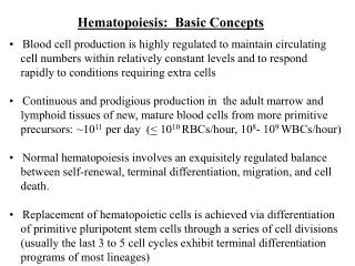
Production of blood cells – bone marrow Blood cells develop in the bone marrow from a common precursor. Once mature, these cells leave the bone marrow and enter the bloodstream, and then the tissues (note brackets at the bottom). We have already looked at these mature blood cells in the bloodstream. As you can see, development of mature blood cells from a common precursor in the bone marrow is quite complicated (and this figure doesn’t show most of the details). Fortunately, we will put this off until the Hematology course in the Fall.
Production of blood cells – bone marrow Just so you’re not left totally in the dark… This is a section of red bone marrow. The nucleated cells are developing blood cells in various stages of development. A = adipose M = megakaryocyte ( produces platelets) Ey = erythrocytes E = developing eosinophils BN = developing neutrophils I repeat, this is all FYI…..for now.
Production of blood cells – THymus This bone marrow box only shows lymphocytes; obviously, other WBCs, RBCs, platelets are developing here too. Most blood cells are mature when they are released into the blood from the bone marrow. The notable exception to this are cells of the T cell lineage, which undergo further maturation in the thymus. This is a complicated and not well understood selection process that involves removal of T cells that would bind to self antigen if released into the general circulation. The details are beyond the scope of this module, but will be discussed in class. In blood smears, T cells that are in transit from the bone marrow to the thymus are indistinguishable from mature T cells that have passed through the thymus, as well as from other non-T cell lymphocytes (e.g. B cells). However, functionally, these cells are immature and will not initiate an immune response.
Production of blood cells – THymus Within the oral cavity, there are lateral outpouchings referred to as the pharyngeal pouches (more on these next year). These spaces are lined by epithelial cells, surrounded by connective tissue. The thymus develops from the 3rd pharyngeal pouches, which pinch off from the oral cavity, and migrates into the neck, anterior to the trachea where they fuse into a single organ. The epithelial cells, called epithelial reticular cells, maintain connections with each other (e.g. desmosomes) to form a network, the “lumen” of which becomes occupied by T cells from the bone marrow. Epithelial reticular cells not only provide the blood-thymus barrier (a protected environment that appears necessary for T cell maturation), but are involved in the antigen-presentation necessary for T cell selection. These epithelial cords are surrounded by connective tissue that forms a capsule around the entire organ, as well as partitions that divide the epithelial cords into lobules. Blood vessels reach the thymus through these connective tissue elements. tongue
Production of blood cells – THymus cortex medulla The thymus is surrounded by a connective tissue capsule (blue arrows, thickness varies due to tissue removal). Extensions of the capsule, called trabeculae (yellow arrows), partition the thymus into lobules (yellow outline) that vary in size, though some of this variation is due to sectioning. Each lobule has a darker outer portion, the cortex, containing more immature T cells, and a lighter medulla where more mature T cells are located. The trabeculae do not penetrate through the entire organ, so that the lobules are not completely separate structures. The medulla is a single, convoluted structure, the extensions of which are covered by cortical tissue.
Production of blood cells - thymus Video of overview of the thymus – SL81 • Link to SL 081 • Be able to identify: • Thymus • Capsule • Trabecula • Cortex • Medulla • Lobules
Production of blood cells – THymus cortex medulla cortex cortex cortex A medium power view of a lobule, showing the cortex and medulla. Recall from the connective tissue and blood (inflammation) labs that when there are white blood cells in a region, the clustering of numerous nuclei of these cells creates a darker basophilic region. In this case, T cells are more closely packed in the cortex relative to the medulla, giving it a darker stain.
Production of blood cells – THymus cortex medulla In this high powered image from the thymus, the cortex and medulla are indicated. The numerous small nuclei are T lymphocytes, which are more numerous in the cortex. Epithelial reticular cells (black arrow) are seen more easily in the medulla, characterized by a large, euchromatic nucleus and abundant, pale-eosinophilic cytoplasm. Many of these cells have begun to cluster together in a concentric arrangement to form structures called Hassall’s corpuscles (yellow outline), a characteristic feature of the thymus.
Production of blood cells - thymus Video of details of the thymus – SL81 • Link to SL 081 • Be able to identify: • Thymus • Lymphocytes • Epithelial reticular cells • Hassall’s corpuscles
Aging of the THymus Premature baby 4-year-old child You won’t have to worry about dating the thymus. (In fact, some of you already have a significant other.) Furthermore, since the aged thymus is largely replaced by adipose tissue, it would be difficult to definitively identify an adult thymus. 17-year-old 36-year-old As the thymus ages, it undergoes histological changes. Continued formation of Hassal’s corpuscles (black arrows), and an increase in the size of these structures, occurs throughout early childhood and adolescence. These corpuscles undergo “keratinization”, so that larger corpuscles have little cellularity. During adult life, the thymus undergoes involution, being replaced by adipose tissue, until it no longer resembles the thymus at all. “Keratinization”? Yes, like the keratinization in stratified squamous keratinized epithelium.
Production of blood cells Video of the thymus of a premature baby – SL81 Video of the thymus of a 3-year-old – SL80 Video of the thymus of a 17-year-old – SL82 Video of the thymus of a 36-year-old – SL34 • Link to SL 081and SL 080and SL 082and SL 034 • Be able to identify: • Thymus • Nothing new that is specific, just appreciate the changes in the thymus during aging
The next set of slides is a quiz for this module. You should review the structures covered in this module, and try to visualize each of these in light and electron micrographs. • Distinguish, at the light microscope level, each of the following:: • (Bone Marrow) • Thymus • Capsule • Trabeculae • Cortex • Medulla • Lobules • Hassal’s corpuscles • Epithelial reticular cells • Lymphocytes • Distinguish, in electron micrographs, each of the following : • Be able to interpret electron micrographs of lymphoid organs if given the source tissue
Final quiz Self-check: Identify all structures. (advance slide for answer) Unilocular adipose trabeculae capsule Hassell’s corpuscle
Final quiz Self-check: Identify the regions. (advance slide for answer) cortex medulla
Final quiz Self-check: Identify the cells. (advance slide for answer) Lymphocytes (T cells) Epithelial reticular cells (these are awesome!!!)
Final quiz Self-check: Identify the organ and region. (advance slide for answer) thymus lobule
OK, this quiz feels pretty pointless right now. Let’s wait until we have more immune organs to confuse you with.