Download
1 / 19
200 likes | 354 Views
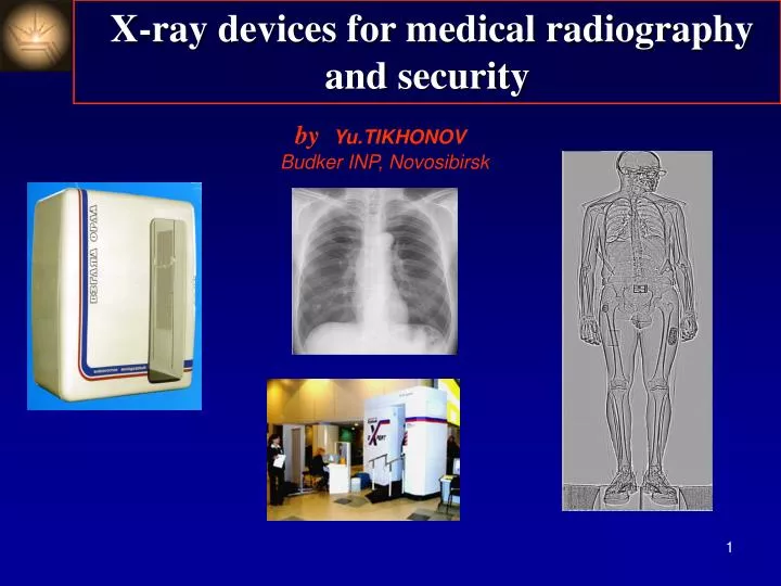
X-ray devices for medical radiography and security. by Yu.TIKHONOV Budker INP, Novosibirsk. During last years the new low dose X-ray devices for medical and security applications have been developed

E N D
X-ray devices for medical radiography and security by Yu.TIKHONOV Budker INP, Novosibirsk
During last years the new low dose X-ray devices for medical and security applications have been developed • in Budker INP • Why we need low (micro) dose technology? • Medicine: • -situation with tuberculosis and cancer requires the mass • inspection of population (at least once per year) • -present fluorography gives very high dosesrisk of cancer! • Security: • -due to problems with terrorism the detection of dangerous • items and weapons hidden on body (and inside the body too) • is very important. • -unlike inspecting for metal items, radiographic inspection is • the only way to inspect for plastic explosive materials and • weapons. • -these devices can be used at airports, customs, prisons, • embassies, nuclear power centers, banks etc.
How to obtain low dose? • Minimization the scattering radiation (scanning systems!) • High efficiency detector • Low noise detector • “Good” X-ray optic • In this case the dose will be limited only by statistics!
Scanning method vs 2D imaging 2D SCAN 2D
Scanning method vs 2D imaging • Advantagesof scanning method • the dose ~10 times lower at same contrast (depends on thickness) • no artefacts • the image length is not limited (up to 2 m) • good image quality in hole field • big dynamic range • Disdvantages • long scanning time (3-5 s vs 0.01 sec in 2D methods) • precision mechanics • One remark:it seems in the case of scanning method an imageis not sharp due to movements of internals but it is not true! The time for read out of one string is very short (<0.001 s) and image dynamically is very sharp and provide additional information for a doctor.
MIC schematic diagram: 1 – detector housing; 2 – anode plane; 3 – cathode strip plane; 4 – input diaphragm; 5 – readout electronics. High efficency and low noise detector: Multichannel Ionization Chamber (MIC) Xe, Kr up to 40 bar (Reticon and Indigo chips are used for readout) length*pressure
Multichannel Ionization Chambers (MIC) A few types of MIC’s have been developed for different applications Efficiency of MIC’s: 0.60.8
Multichannel Ionization Chambers (MIC) • Advantages of MIC : • Low noise • Large dynamic range • High radiation resistance • Fill factor 100% • Disadvantage: • High pressure The MIC800 for security (800 mm)
The devices for medical application based on MIC Fluorograph DRC South Korea Fluorograph FMC – NP –О Russia
Some parameters of FMC-NP-o (Russia) -------------------------------------------------------------------------- Image sizes, mm410 х 1200 Resolution, pl/mm2 Contrast sensivity , %0,5 (at dose 100 Rin detector plane) Scan speed , cм/s10 Dynamic range 1000 Dose for chest image , Зv5 HV on X-ray tube (max), кV120 Distance: X-ray tubedetector, mm 1300 # images/hour 60 ------------------------------------------------------------------------- Four companies (2 in Russia, 1 in China and 1 in Korea) produce medical devices based on MIC’s detectors by license of BINP .
Application for security System for Radiographic Control (SRC) Requirements
Column Detector X-ray beam Irradiator Radiolucent stand Motor A principal design for SRC
Basic SRC parameters The permitted years dose* (in many countries) is 1000 Sv Each passenger can pass SRC inspection many times without any trouble *medical inspections are not included!
Dose levels (Зв) 1. Medical inspections: • Computer tomograph 10 000 • Chest inspection, mamography novel digital devices 30-100 novelfilm X-ray200 2. Radiation background: • At 1500 msee level (per day) 6 • At 10000 m (per hour) 5 3.SRC inspection <0.5
SRC installed in Tolmachevo airport in Rusiia Up to now 6 SRC devices are in operation in four Russian airports. Welcome!
Conclusion • The set of Multichannel Ionization Chamber’s (MIC) have been • developed for practical usage in medicine and security • The scanning devices based on MIC’s allow to have lowest doses • compare to other methods • A few years of operations of the different devices based on MIC’s • shown good efficiency and reliability • The new developments are in progress: • the goal -decreasing of the scanning time (electronics+mechanics)