Download
1 / 7
70 likes | 90 Views
QIN Informatics and Software Tool Sharing 3D Slicer. Andriy Fedorov, Brigham and Women’s Hospital fedorov@bwh.harvard.edu.

E N D
QIN Informatics and Software Tool Sharing3D Slicer • Andriy Fedorov, Brigham and Women’s Hospital • fedorov@bwh.harvard.edu
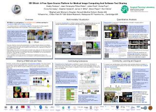
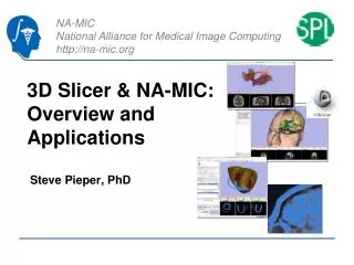
Visualization of multi-parametric prostate MRI dataset that includes dynamic contrast-enhanced (DCE) DWI and T2-weighted MRI. Interactive plotting of the signal intensity over time is provided by Slicer MultiVolume support infrastructure. DCE MRI pharmacokinetic modeling functionality is currently under development and will be available as Slicer extension. Visualization of a multi-modal brain tumor resection planning dataset. 3D visualization includes surface model of the tumor and ventricles boundary, fiber tracks, and fiducial annotations. Overview • Multi-modality visualization, Segmentation, Registration • Free open source with no strings attached • Binary packages for Win, Mac and Linux • Community and user support • Research software, not FDA approved • http://slicer.org
Slicer Extensions • Lean core application + core modules • Extensions • same flexibility as core modules • do not need to adhere to Slicer license • “Application store” concept
SparKit is a project funded by An Applied Cancer Research Unit of Cancer Care Ontario with funds provided by the Ministry of Health and Long-Term Care and the Ontario Consortium for Adaptive Interventions in Radiation Oncology (OCAIRO) to provide free, open-source toolset for radiotherapy and related image-guided interventions. Pinter C, Lasso A, Wang A, Jaffray D, Fichtinger G. SlicerRT: radiation therapy research toolkit for 3D Slicer. Medical physics. 2012 October;39(10):6332–8. SlicerRTOntario Consortium for Adaptive Interventions in Radiation Oncology (OCAIRO)
iGyneTina Kapur, Akila Viswanathan, Brigham and Women’s Hospital P41 EB 015898 National Center for Image Guided Therapy (Jolesz, Tempany) 2005-2015, R03 EB 013792 Segmentation for Gynecologic Brachytherapy (Kapur) 2011-2013 Kapur T, Egger J, Damato A, Schmidt EJ, Viswanathan AN. 3-T MR-guided brachytherapy for gynecologic malignancies. Magnetic resonance imaging. 2012 November;30(9):1279–90.
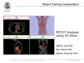
Longitudinal PET/CT analysis extension (shown) integrates ROI segmentation, PET Standard Uptake Value (SUV) quantification and versatile visualization of multiple time-points into a single processing workflow. P.Mercea, A.Fedorov, S.Pieper, R.Beichel, M.-A.Park, J.Hainer, M.F.Kijewski, L.Horky, R.Kikinis, H.Dickhaus "Quantification of longitudinal tumor changes using PET imaging in 3D Slicer" In Proc. Computer Assisted Radiology and Surgery, 27th International Congress and Exhibition, 2013 (accepted) Longitudinal PET/CTP.Mercea (U.Heidelberg), A.Fedorov, S.Pieper, R.Kikinis (BWH), R.Beichel (U.Iowa)