Download
1 / 44
440 likes | 821 Views
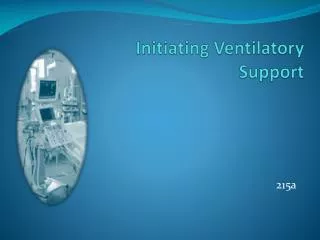
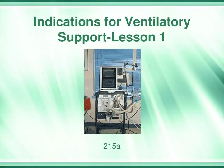
Indications for Ventilatory Support-Lesson 1. 215a. Educational Objectives. Lung Mechanics review List the primary indications for the initiation of ventilatory support Define respiratory failure (type I and II) List the absolute contraindication to mechanical ventilation

E N D
Educational Objectives • Lung Mechanics review • List the primary indications for the initiation of ventilatory support • Define respiratory failure (type I and II) • List the absolute contraindication to mechanical ventilation • List the relative contraindications to mechanical ventilation
transairway pressure transrespiratory pressure transthoracic pressure volume elastance = Dpressure / Dvolume Lung Mechanics resistance = Dpressure / Dflow flow
Respiratory System Review • http://www.youtube.com/watch?v=ka-3qsN3v-E • Review on elastance and compliance
Static Compliance • Cs = tidal volumecorrected for gas compression Pplat – PEEP total peep Normal 100 - 200 ml / cmH2O (PDQ) Decreased with: • Mainstem Intubation • Congestive Heart Failure • ARDS • Atelectasis • Consolidation • Fibrosis • Hyperinflation • Tension Pneumothorax • Pleural Effusion • Abdominal Distension • Chest Wall Edema • Thoracic Deformity
Principle #1: Ventilation The goal of ventilation is to facilitate CO2 release and maintain a normal PaCO2 • Minute Ventilation (Ve) • Total amount of gas exhaled per minute • Ve = Vt x f • Ve comprised of 2 factors • VA = alveolar ventilation • VD = dead space ventilation • Ventilation in the ICU setting • Increased CO2 production • Fever, sepsis, injury, overfeeding • Increased VD • Vent circuit, ET tube • Adjustments: Vt and f
Fig. 13-1. Factors that affect the partial pressure of arterial carbon dioxide (PaCO2) during mechanical ventilation. V.CO2, carbon dioxide production; V.A, alveolar ventilation; V.E, minute ventilation; V.D, dead space ventilation; VT, tidal volume; TI, inspiratory time; TE, expiratory time; f, respiratory rate. (From Hess DR, MacIntyre NR, Mishoe SC, et al: Respiratory care principles and practice, Philadelphia, 2002, WB Saunders.)
Principle #2: Oxygenation The primary goal of oxygenation is to maximize O2 delivery to the blood (PaO2) • Alveolar-arterial O2 gradient • Equilibrium between O2 in the blood and O2 in the alveoli • A-a gradient measures efficiency of oxygenation • PaO2 partially depends on ventilation but more on V/Q matching • Oxygenation in the ICU setting • PaO2/PAO2 ratio (a/A ratio), PaO2/FIO2 (>400) (less 200= ALI) • Indicator of efficiency of O2 transport • CaO2 • Adjustments: FiO2 and PEEP
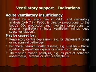
Respiratory Failure What is respiratory failure? Inadequate gas exchange by the respiratory system, with the result that arterial oxygen and/or carbon dioxide levels cannot be maintained within their normal ranges.. Acute ventilatory failure: Defined by an acute rise in PaCO2 and respiratory acidosis (pH<7.25). PaCO2 is directly proportional to the body‘s CO2 production and inversely proportional to alveolar ventilation (minute ventilation minus dead space ventilation).
Primary indications for the initiation of ventilatory support • Type I: Hypoxic Respiratory failure w/o Hypercapnia • Type II: Ventilation Failure w/ hypercapnia and hypoxemia. Can be: • Acute • Impending • Acute on Chronic
Type I Respiratory Failure • Type 1 respiratory failure is defined as hypoxia without hypercarbia, and indeed the PaCO2 may be normal or low. It is typically caused by a ventilation/perfusion (V/Q) mismatch; the volume of air flowing in and out of the lungs is not matched with the flow of blood to the lungs. • The basic defect in type 1 respiratory failure is failure of oxygenation characterized by: • PaO2 low (mod/severe hypoxemia) PaCO2 normal or low PA-aO2 increased
Type I Respiratory Failure • Severe hypoxemia – PaO2 < 40 mmHg • An A-a gradient of greater than 350 while breathing 100% oxygen • Clinical indications of hypoxemic ventilatory failure • Increase in A-a gradient (PAO2 – PaO2) • Increase in A-a ratio (PaO2/PAO2) • PaO2 of 60 mmHg or less from arterial blood gas sample
Type I Respiratory Failure • Severe hypoxemia • PaO2< 60 mmHg on an FIO2 > 0.50 (V/Q mismatching/Shunt = need for mechanical ventilation) • P(A-a)O2 ≈ 306 assuming normal PaCO2 and respiratory quotient
Type I Respiratory Failure • Causes of hypoxemic respiratory failure • Atelectasis • Pulmonary edema • Acute respiratory distress syndrome (ARDS) • Lung injury • Cardiac failure/poor perfusion/increased VO, decreased oxygen carrying capacity, right/left shifts in O2 curve • Pulmomary Emboli • http://www.youtube.com/watch?v=GJLWwl1Z03k&feature=related
Type I Respiratory Failure • http://www.youtube.com/watch?v=VIQC_tCHBBI
Ventilation w/o perfusion Ventilation in near equal parts as perfusion Perfusion w/o ventilation
Type II Respiratory Failure • Acute ventilatory failure • Sudden (Acute) increase in PaCO2with accompanying respiratory acidosis • Presence of cyanosis and/or apnea • Mild to moderate hypoxemia alone does not define acute ventilatory failure • Ventilation = PaCO2 Oxygenation = PaO2
Type II Respiratory Failure • Causes • Increase in dead space (from bronchospasm) • Increase in production of carbon dioxide (infection/Sepsis) • Decrease in alveolar ventilation (atelectasis/pneumo…)
Type II Respiratory Failure • Physiological underlying factors • Abnormal response of respiratory center of brain to changes in PaCO2 (brain injury/bleeding, spinal injury) • Neurologic impulses from respiratory center of brain not reaching respiratory muscles (Neuromuscular problems) • Respiratory muscles and lungs unable to respond to neurologic impulses
Type II Respiratory Failure • Examples of disease states leading to acute ventilatory failure • Acute exacerbation of COPD (acute on chronic, pneumonia/infection…) • Tumor of brain or nervous system (leading to impairment of PONS) • Trauma to head (bleeding…)
Type II Respiratory Failure • Examples of disease states leading to acute ventilatory failure • Pneumonia (post op, acute/aquired…) • Pulmonary edema (cardiogenic/non-cardiogenic) • Cerebral vascular accident (stroke/ ischemic/ hemorrhage)
Chronic to Acute Ventilatory Failure • Examples of disease states leading to acute ventilatory failure • Guillain-Barre syndrome • Myasthenia gravis • Poliomyelitis • COPD/Asthma with an acute exacerbation
Increased Production on Co2 • Infection • Sepsis • Burns • Metabolic Alkalosis
Type II Respiratory Failure-impending failure • Impending ventilatory failure • Patient able to maintain marginally normal blood gases, but only with a significantly increased work of breathing • PaCO2 may be normal or decreased during this period • Patient with increased WOB/accessory muscle usage
Type II Respiratory Failure-impending failure • Cause • Respiratory muscle fatigue or weakness • Decrease in capacity of rested muscle to generate force and decrease in endurance of muscle, patients appear to be “tiring out”
Type II Respiratory Failure-impending failure • Respiratory muscle fatigue or weakness (neuromuscular diseases) • Assessment of muscle strength • Maximum inspiratory pressure (MIP) normal -80 cmH2O, intubate at -20 cmH2O; measures ability to take a breath in • Maximum expiratory pressure (MEP) normal 100-150 cmH2O, measures ability to cough • Forced vital capacity (FVC); a MIP and MEP combined • Maximum voluntary ventilation (MVV), a FVC performed over about 15-20 seconds
MIP (NIF) and MEP Maneuver can be done digitally or through a Wright Respirameter http://www.youtube.com/watch?v=Cnbh2i74FaI
Impending Ventilatory Failure • Causes of muscle fatigue • Excessive demand placed on muscle • Hypoxemia • Decreased blood flow to the muscle (perfusion problems) • Poor nutrition (homeless, alcoholics, elderly…) • Inability of muscle to extract nutrients from blood
Impending Ventilatory Failure • Examples of disease states leading to muscle weakness or fatigue • Neuromuscular diseases • Chronic obstructive pulmonary disease • Morbid obesity (CO2 retention) • Kyphoscoliosis (secretion clearance problems)
Impending Ventilatory Failure • Work of breathing • Amount of pressure needed to move a given volume of gas into the lung with a relaxed chest wall • Most common cause of muscle fatigue • Composed of two forces • Physiologic work • Effort required to overcome elastic forces during inspiration plus the airway resistance and lung tissue resistance • Increased in both airway and parenchymal abnormalities • http://www.youtube.com/watch?v=TKyoEmA3bGE
Impending Ventilatory Failure • Work of breathing • Composed of two forces • Imposed work • Effort required to overcome airway resistance of artificial airways, ventilator tubing, and auto-PEEP • Can be an impediment to weaning
Airway Resistance (Change in Pressure/Flow) • Airway resistance is the opposition to flow caused by the forces of friction. It is defined as the ratio of driving pressure to the rate of air flow. Resistance to flow in the airways depends on whether the flow is laminar or turbulent, on the dimensions of the airway, and on the viscosity of the gas. • The most important variable is the radius, which, by virtue of its elevation to the fourth power, has a tremendous impact on the resistance. Thus, if the diameter of a tube is doubled, resistance will drop by a factor of sixteen. • For turbulent flow, resistance is relatively large. That is, compared with laminar flow, a much larger driving pressure would be required to produce the same flow rate. Because the pressure-flow relationship ceases to be linear during turbulent flow, no neat equation exists to compute its resistance. • Resistance caused by: Narrowed airway
Impending Ventilatory Failure • Clinical indications of impending ventilatory failure • Decreasing tidal volume • Increasing respiratory rate • Labored or irregular breathing pattern • Increasing PaCO2 over serial blood gases • Tachycardia
Prophylactic ventilatory support • Prophylactic ventilatory support • Pre surgical patients • Aspiration risks (CVA, drug OD…) • Risk of pulmonary complications, ventilatory failure, or oxygenation failure is high • Minimizes hypoxia of major organ systems, promoting patient recovery • Prolonged shock • Head injury • Smoke inhalation
Prophylactic Ventilatory Support • Indications • Hypoxia of brain, myocardium, or other major organ tissue • Coronary artery bypass surgery • Thoracic or abdominal surgery
Prophylactic Ventilatory Support • Support provided when risk of pulmonary complications, ventilatory failure, or oxygenation failure is increased
Contraindications to Ventilatory Therapy • Absolute contraindication • Untreated pneumothorax Heart and trachea deviated to right
Contraindications to Ventilatory Therapy • Relative contraindications • Patient’s informed request (DNR, modified code) • Medical futility – if intervention was useless in the last one hundred cases of the physician, it is reasonable to believe that intervention would again be futile
Contraindications to Ventilatory Therapy • Relative contraindications • Reduction or termination of patient’s pain and suffering • In cases of terminal disease, the benefit of ventilation must be weighed against the degree of pain and length of suffering which the procedure would engender