Download
1 / 57
610 likes | 686 Views
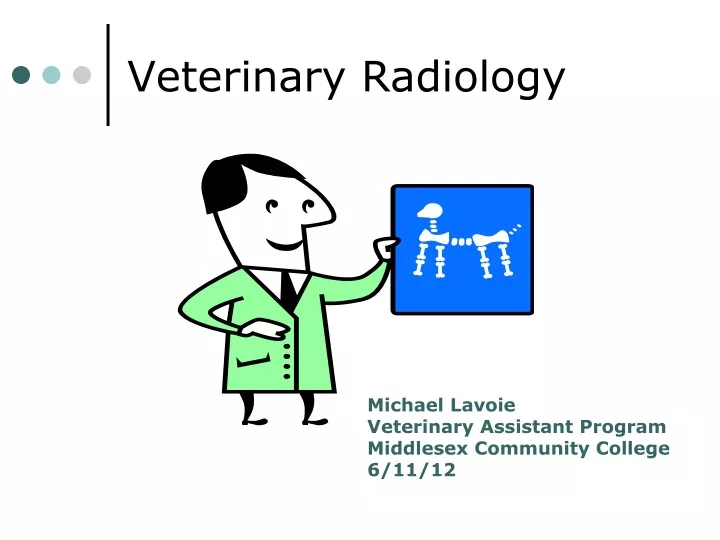
Veterinary Radiology. Michael Lavoie Veterinary Assistant Program Middlesex Community College 6/11/12. You will learn…. The basic concepts of Veterinary Radiology How the technology of radiology has advanced The benefits animals gain from the advancements of radiology

E N D
Veterinary Radiology Michael Lavoie Veterinary Assistant Program Middlesex Community College 6/11/12
You will learn…. • The basic concepts of Veterinary Radiology • How the technology of radiology has advanced • The benefits animals gain from the advancements of radiology • Why safety is important while being involved with radiology • The different careers involved within Veterinary Radiology
The Basics • Radiology is used to see injuries, fetuses, diseases, abnormalities, and damages done to internal body parts and organs. • For the most part, radiology provides a quick diagnosis in most emergency situations. • Radiology is used everyday with clinics. • Has advantages and disadvantages.
The Basics • Radiology is very beneficial. • Without radiology, surgery would have to be fulfilled to diagnose problems within the body of an animal. • Radiology also helps to expand the information obtained from clinical examinations of animals.
The Advancements of Veterinary Radiology Technologies Advantages Disadvantages
Digital Radiology • Higher dynamic range than film • Provides more information at high and low exposures
Digital vs. computed vs. Direct Capture radiology • Computed radiology • Uses a cassette with an imaging plate • Digital radiology • Uses an linear array of radiation detectors • Similar to computed radiology • Direct capture • Uses a solid state radiation device.
Computed Radiology • A cassette is used • Contains a photostimulable phosphor imaging plate • Looks like an intensifying plate • When struck with x-rays, a new semi-stable state is produced. • The latent image is stored on the plate
Imaging plate • Made up of multiple layers • A polyester support layer • A phosphor layer • A clear protective layer • Conductive layer • Support layer • Backing layer • Bar code layer on the back • Contains a number assigned to the image plate.
Imaging plate • Very flexible • Can maintain a latent image for about 24 hours • Can be exposed to light • Some image degregation may occur with extended storage time and light exposure
Terminology • Rotation/inversion • The ability to change the image presentation, or turn the image from a negative to a positive • Anatomic measurements • The ability to measure certain areas of interest • Short-term database functions • Allows user to locate images, create lists if images, image interpretation, and maintain teaching files
The Veterinary Direct Radiology System (Vet DRS) • Developed within the past ten years. • The Vet DRS was created to preserve space and provide a quick diagnosis. • This system is noted for its ability to store many radiographs as well as produce accurate radiographic images that provide for an accurate diagnosis. • Designed in both a small and large arrangement.
The Veterinary Direct Radiology System (Vet DRS) • Smaller arrangements are portable and were made for the use of field vets. • It was made for the vet to take shots of things such as a horses leg during the time of emergency for a quick diagnosis without being exposed to the radiation beams. • Images appear instantly appear onto the screen of a laptop carried by the vet to provide an on the spot diagnosis.
The Veterinary Direct Radiology System (Vet DRS) • Larger arrangements are not portable. • They remain within a radiology suite inside a veterinary clinic • The Images instantly appear onto a monitor to be reviewed by the medical staff • All arrangements are digital.
Digital Radiology • Does not use cassettes • A radiation detector array is used • These detectors are gas filled • The more detectors, the better the spacial recognition
Digital Fluoroscopy • A computer and monitor are needed • The monitor is used to enter and edit patient data and to display the image. • Image is displayed immediately and can be modified and changed almost immediately as well.
PACS • Picture Archive and Communication System • Allows for the digitalization of conventional radiographs • Allows for acquisition, interpretation, and storage of images.
Three components of PACS • Display system • Network • Storage system
Protocols created for use on horses • One certain advancements allow vets the ability to produce accurate images of a horse’s legs. • “Magnetic Resonance Imaging” (MRI) • An MRI can now be taken on a horses leg. • Before this was developed surgery would have been the only way to determine the damage of a horse’s leg.
Protocols created for use on horses • Bone diseases or abnormalities, ligaments, tendons, and joints can be examined by this technology. • Color can be added and images can be made 3-D using this system. • Allows a realistic image of the inside of a horse’s leg.
Computers • Computers now contribute to the field of Veterinary Radiology. • Most radiographs are now stored within computers to Also they provide clearer preserve space. • images and certain programs to make radiographs seem more realistic and easier to understand.
Advantages of the Advancements • The advancements provide quicker and more reliable images and diagnosis. • They also allow the creation of more effective treatment plans.
Disadvantages • The only disadvantage of some advancements is that some radiographs may be misinterpreted or taken wrong. • This results in the taking of more radiographs which causes a prolonged diagnosis.
The Dangers and effects • Scatter radiation can cause cellular damage which is basically the destruction of the cells within the body and may result in cancer or another hazardous illness
Regulations • Individuals under the age of 18 may not be involved in any type of radiological activity because they have not fully developed; therefore, they are efficiently harmed by radiation exposure. • Pregnant women should also not be involved within radiological activities because their developing fetus will be easily harmed by the least amount of radiation expossure.
The Order of Operations • Created to aid in the protection of both humans and animals against the hazardous “scatter radiation”. • The Order of Operations consist of the following: an adequate technique chart or comparable system, positioning aids, protective clothing and other protective barriers, personnel dosimetry devices, emergency procedures for malfunctioning x-ray equipment, and quality control measurements and tests.
Animal Safety • Not as vital as it may be for humans • However, reproductive organs must be protected to prevent any type of damage. • Fetuses within a pregnant female may become harmed if the correct safety procedures are not followed. • Lead shields are also available for animals.
Reading Rads (Terms) • Radiolucent: almost entirely transparent • Radio-opaque: not transparent to x-rays • In order from radiolucent to radio-opaque: air, fat, water, bone, metal • “scatter radiation”: appears cloudy on radiographs—scatters throughout the radiology suite—causes more exposure to individuals involved
Case Study 1 • Signalment • 3 year-old female spayed domestic short-haired cat • History • anorexia, coughing, depression for 2 days • Physical Exam • Pink mucous membranes • Crackles ausculted cranioventrally on right side • Respiratory rate was 80 breaths per minute
Is the size/shape of the heart normal? • Yes • Normal Size and shape of a heart
How do the lungs look? • This radiograph shows an abnormal lung pattern.
Does the diaphragm shape and position look normal? • No: • Flattened diaphragm outline.Increased distance between heart and diaphragm.
Bronchial Pulmonary Pattern • Characteristics:Increased radiopacity.Opacities centered around bronchi ("donuts")Diffuse, non-homogenous.Pulmonary vessel margins indistinct.Prominent longitudinal peribronchialopacities.These findings are characteristic ofa bronchial pulmonary pattern.
Case Study 2 • Signalment • 13 year-old female spayed mixed breed dog. • History • Dog presented with a two week history of anorexia, lethargy and polyarthritis. • Physical Exam • The carpi and tarsi were warm and swollen.
What are some changes you have noted? • There are multiple abnormalities presentinvolving the carpal region: • Scalloped-type periosteal proliferationinvolving primarily abaxial and dorsal aspects of the second and fifth metacarpal.Similiar periosteal proliferativechanges of radius and ulna.Soft tissue swelling.No joint involvement.Multiple bones involved.No lysis of the cortical cavity.
What radiographs would you take? • Chest? • Abdominal? • Opposite carpus? • Tarsi? CHEST!
What is the diagnoses? • Pulmonary mass: Characteristic of a nodularinterstitial pulmonary pattern. • These masses represent eithera primary pulmonary neoplasia ora metastatic pulmonary disease.
Case Study 3 • Signalment • 2 year-old female domestic shorthair. • History • Cat vomiting for 3 days. • Physical Exam • Nothing abnormal was noted on abdominal palpation
Are the kidneys normal size? Yes! They are normal size and shape!
What is this? • There is mild gas and fluid distensionof the descending intestine.
Diagnoses? • The small intestinesare "bunched" into the mid-abdomenon the survey radiographs.This is commonly found in obese catsand must be distinguishedfrom abnormal "bunching" thatoccurs with linear foreign bodies.In the case of linear foreign bodies,there usually will be an abnormalcurvature of the involved loops("hairpin" turns) and someexcess gas within the lumen.