Download
1 / 41
420 likes | 442 Views
Neurocritical care for retrieval medicine. Stuart Lane. Overview. Traumatic Brain Injury TBI Subarachnoid haemorrhage SAH Arteriovenous Malformation AVM Intracranial Haemorrhage ICH Ischaemic stroke Specific therapies e.g. vasospasm Monitoring and drains Extras. TBI.

E N D
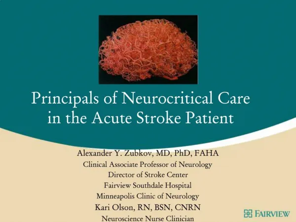
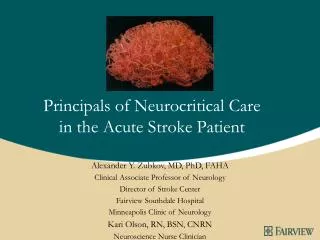
Neurocritical care for retrieval medicine Stuart Lane
Overview • Traumatic Brain Injury TBI • Subarachnoid haemorrhage SAH • Arteriovenous Malformation AVM • Intracranial Haemorrhage ICH • Ischaemic stroke • Specific therapies e.g. vasospasm • Monitoring and drains • Extras
TBI • The leading cause of death and morbidity from ages 1- 44yrs old • The latest Brain Trauma Foundation (BTF) guidelines 2007 are well accepted • Prehospital • Hypoxaemia and hypotension • GCS used in pre-hospital setting DISCUSS GCS • Non-use paralysis for intubation • Hypertonic NaCl for GCS <8 can be considered • Hyperventilation for signs of herniation • Penetrating injury also included • Initial medical management no different • Surgical management different • Most likely to be blunt closed TBI
Interhospital transfer • Avoid hypotension and hypoxaemia • Hypotension is worse than hypoxaemia • Dramatically worsen outcome • Intubation is assumed for this presentation • Oxygen PaO2 > 90mmHg / SpO2 > 95% • PaCO2 35-40 mmHg / PETCO2 31-35 mmHg • CPP cant be assessed without an ICP monitor • MAP > 8O mmHg if no monitor • CPP 50-70 if monitor. • What do we mean by CPP? • What did the trials really show / ?ARDS
Interhospital transfer • Phenytoin only if witnessed seizure • Resuscitation to euvolaemia with isotonic crystalloids, then use noradrenaline to augment MAP FLUIDS • NGT / OGT • Normothermic • If spine cleared, then sit 30° head up • Brown tape vs.. white tape • Remove collar and use sandbags – venous congestion • Avoid hyperglycaemia – not really a retrieval issue • Paralyse for transfer • No evidence of improving ICP • Significant amount of movement for retrieval • BTF not in retrieval patients • Paralysis not evidenced, not worried about CIPMN at this time • May make the patient more CVS stable
ICP monitoring and EVD’s • The transducer is fixed at a reference level • Foramen magnum / external auditory meatus. • The system is connected to a drainage chamber • Allows drainage of CSF into the collecting bag or chamber. • The height of this drainage chamber can be adjusted relative to the reference point • When the EVD is unclamped and the stopcock is opened it allows drainage of CSF • When the ICP is more than the set height of the draining chamber. • The drainage will continue until • The ICP falls to a value less than the chamber height • The EVD is clamped.
ICP monitoring and EVD’s • P1- Percussion wave • Created by systolic blood pulsation transmitted through choroid plexus) • P2- Tidal wave • Reflects brain compliance • Exceeds P1 in noncompliant brain • P3- Dicrotic wave • Produced due to aortic valve closure
ICP monitoring and EVD’s • A waves - plateau waves • Lasting 5–20 min • 50 –100 mmHg high • B wave • Frequency of 0.5–2 min-1 • Up to 50mmHg • C waves • Last 4–5 min-1 • Up to 20mmHg • Intervention is required for A and B waves • C waves may be • Normal • Due to a change in the vasomotor tone.
Closed vs open EVD • Limit clamping of EVD’s • Administration of intrathecal antibiotics • Assessment for removal • Getting the patient into a chair / back into bed • Clamp it whilst transferring the patient from bed to stretcher • Avoid loss of csf if the drain goes below the level of required drainage • Open it again and leave open for transfer • For a long transfer, if known ICP problems, then measure ICP and check pupils at intervals • Clamp it again when transferring the patient from stretcher to bed
What if the ICP goes up • Recheck everything • ABC and glucose • Monitoring equipment including waveform • Deepen sedation • Paralyse, if not already done • Deepen sedation further • Consider CSF drainage / open drainage system • Osmotherapy • Mannitol vs. hypertonic saline • Manual hyperventilation in an emergency
Second line ICP therapy • ABC again • Recheck the waveform if you have one • If ICP remains > 20 mmHg for 15 minutes despite the above treatment • More sedation • Optimise fluids. • Consider diuresis • EVD left open • Mild hypothermia • More paralysis • Thiopentone bolus • Maintain MAP with noradrenaline
Forget about.. • Transcranial Doppler • Jugular bulb monitoring • Brain oxygen content monitor • Cerebral microdialysis • Corticosteroids
SAH • 5% of all strokes • 10–15% die before reaching hospital • Pathophysiology • 80% aneurysmal, • 15% AVM • 5% other e.g. clotting abnormalities / drugs • Rebleeding risk • 8% in the first 48 hours • 1% per day thereafter • Reasonable to delay surgery • Poor grade • Established vasospasm
Aneurysmal SAH • Management different if aneurysm is secured or unsecured • Much extrapolation of TBI data • Oxygen PaO2 > 90mmHg / SpO2 > 95% • PaCO2 35-40 mmHg / PETCO2 31-35 mmHg • Fluid resuscitation to euvolaemia with isotonic crystalloid • Noradrenaline to maintain MAP >70mmHg • EVD for hydrocephalus ?CLOT DISRUPTION • Debatable for prevention of hydrocephalus • ICP monitoring for severe cases • OGT / NGT
Surgery • Surgery within 48hrs if possible • Clipping vs.. coiling still a big debate • Improved mortality with coiling • Rebleeding slightly higher in coiling group • Clipping still favoured in the US • Discussion between neurosurgeon and neuroradiologist • Can still Transfer to any neurosurgical centre • Coiling favoured • Patients with poor clinical grade • Patients who are medically unstable • In situations where aneurysm location imparts an increased surgical risk • cavernous sinus • basilar tip aneurysms • Small-neck aneurysms in the posterior fossa • Patients with early vasospasm • Cases where the aneurysm lacks a defined surgical neck • Patients with multiple aneurysms in different arterial territories if surgical risk is high • It is falling out of favour once again in some groups
Transfer of aneurysmal SAH • MAP 70 if aneurysm not secured • 30° head up • Keep normothermic • Avoid hyperglycaemia • Nimodipine can wait TRIALS • Magnesium can wait TRIALS • Monitoring and access • CVC and arterial lines discussed later
Vasospasm • Delayed narrowing of the large capacitance vessels at the base of the brain • Poorly understood (DOES IT EXIST) • Normally 7-10 days post SAH • 70% of SAH patients have angiographic evidence • 30% have significant clinical sequelae • Impaired autoregulation • Cerebral ischaemia • Cerebral infarction • Prevention and treatment are different entities • Prevention • Nimodipine • Magnesium • No other agents • HHH therapy is therapy not treatment • Illogical • Not supported
Vasospasm • Treatment • Fluids to euvolaemia • Try to avoid excess hypervolaemia • Imaging to rule out other possible problems • Titrate MAP to neurological improvement with noradrenaline • MAP 90-100 if unconscious • Angiography • Verapamil / papaverine injections • Angioplasty
AVM • The direct connection between the arterial and venous systems supplies a low-resistance shunt for arterial blood and exposes the venous system to abnormally high pressures • Use systolic BP limitations • Lots of sedation and paralysis for transfer
ICH • 10% of all strokes • Surgical intervention becoming less common • Posterior fossa lesions > 3cm • Young patients with lobar haemorrhage • Association with a structural vascular lesion • STITCH trial • Craniotomy for superficial clots (<10mm from the skull) with clinical deterioration • Aspiration for deep clots
Transfer of ICH • Specific guidelines • MAP <130mmHg in chronic hypertension • No use of rVIIa • Extrapolation of TBI data once again • Sedate • Paralyse
Ischaemic stroke • 85% of all strokes • Thrombosis • Embolism • Hypoperfusion • Venous occlusion • Clinical presentation • Decreased conscious level • CT showing • No haemorrhage • No significant MCA territory involvement
Thrombolysis? • Severe neurological impairment with NIH stroke scale >22 • CT evidence of extensive MCA territory infarct • Sulcal effacement • Loss of GW differentiation • Greater than 1/3 MCA territory
NIH stroke scale • Total score out of 44 • Level of consciousness • Best gaze • Visual • Facial palsy • Motor arm • Motor leg • Limb ataxia • Sensory • Language • Dysarthria • Extinction and inattention • If retrieval required • Likely to be greater than 22 • Unable to assess accurately
Thrombolysis • Within the first 3 hours • substantial net benefits for virtually all patients with potentially disabling deficits. • Within 3-4.5 hours • moderate net benefits when applied to all patients with potentially disabling deficits. • MRI of the extent of the infarct core • can likely increase the therapeutic yield of lytic therapy, especially in the 3 to 9 hour window. • Intra-arterial fibrinolytic therapy in 3 to 6 hours • moderate net benefits when applied to all patients with potentially disabling deficits and large artery cerebral thrombotic occlusions. • Based on NINDS 1 and NIND 2 rt-PA trials
For transfer • Have the primary hospital discuss this with the receiving hospital • IV rt-PA • Aspirin if no haemorrhage on CT • Anticoagulation not indicated unless suggestion of venous infarction • Dose of clexane for transfer • Specialist interventional radiology centre for intra-arterial thrombolysis • New techniques
Other possibilities.. • Hemicraniectomy for significant MCA infarct • Treat as for raised ICP • Surgery is definitive treatment • 18-60 years within 48 hours • More likely to be called when thrombolysis has caused haemorrhagic transformation • Cryoprecipitate infusion • Platelet transfusion • Primary centre to arrange pre-arrival
Other conditions • Cerebral tumours • Consider steroids for raised ICP • Mannitol is effective • Cerebral abscess • Consider steroids for raised ICP
Monitoring • Arterial line • Desirable • Not if delaying treatment • Central Venous Catheter • Desirable • For multiple infusions (Not in retrieval medicine) • For catacholamines • CVP measurement is wasting time and useless • Not if delaying treatment • Don’t delay treatment • Heavy sedation, may require catacholamines • Paralysis may help here
Clinical monitoring of possible cerebral herniation • Difficult if sedated / paralysed • Pupils need to be seen • So do not tape • Clinical examination remains paramount • Emergency measures • Hyperventilation • Osmotherapy • Burr hole
Clinical monitoring of possible cerebral herniation • Can occur at ICP’s <25mmHg • ICP threshold is not uniform • Depends on the location of the mass lesion • Abnormal posturing on presentation • Pupillary abnormalities
Brain herniation syndromes • Supratentorial herniation • 1 Uncal • 2 Central (transtentorial) • 3 Cingulate (subfalcine) • 4 Transcalvarial • Infratentorial herniation • 1Upward (upward cerebellar) • 2 Tonsillar (downward cerebellar)
Summary • Keep it simple for transfer • Maintain brain perfusion • Use MAP’s appropriate to pathology • Most specific therapies can wait • Monitoring remains clinical, with assistance from numbers • Avoid delays with primary patholgies